


Ask our dispensing optician
Your request was successfully submitted!

Reviewed by
Beck Jinnette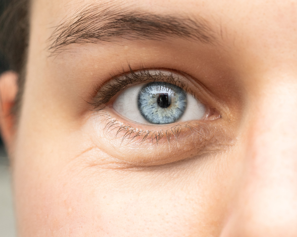
Human beings have five senses, sight being one of them. Eyes are responsible for giving us the sense of vision and allow us to see and experience all the beauty that surrounds us.
One of the most important organs in the body, eyes play a major role in day-to-day life and experiences. “Eyes are the windows to the soul” is an expression that is often used to describe the deep connection we feel when making eye contact with someone.
It is through our eyes and the sense of sight that we are able to connect with people, places and things. Small but mighty, it is essential to keep our eyes healthy. Most people tend to take sight for granted, without giving much thought to how it is all made possible.
Human beings have five senses, sight being one of them. Eyes are responsible for giving us the sense of vision and allow us to see and experience all the beauty that surrounds us.
One of the most important organs in the body, eyes play a major role in day-to-day life and experiences.
“Eyes are the windows to the soul” is an expression that is often used to describe the deep connection we feel when making eye contact with someone. It is through our eyes and the sense of sight that we are able to connect with people, places and things.
Small but mighty, it is essential to keep our eyes healthy. Most people tend to take sight for granted, without giving much thought to how it is all made possible.
What makes up an eye?
The human eye is a complex organ that enables us to perceive the world around us by capturing and processing light. The eye is made up of many light-sensitive cells and works in conjunction with the brain to provide us with the sense of vision.
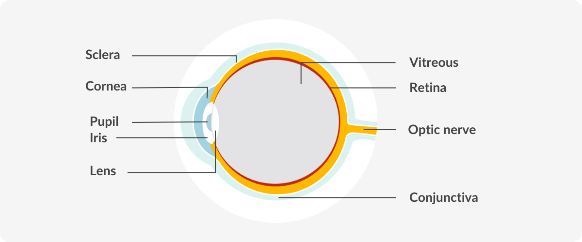
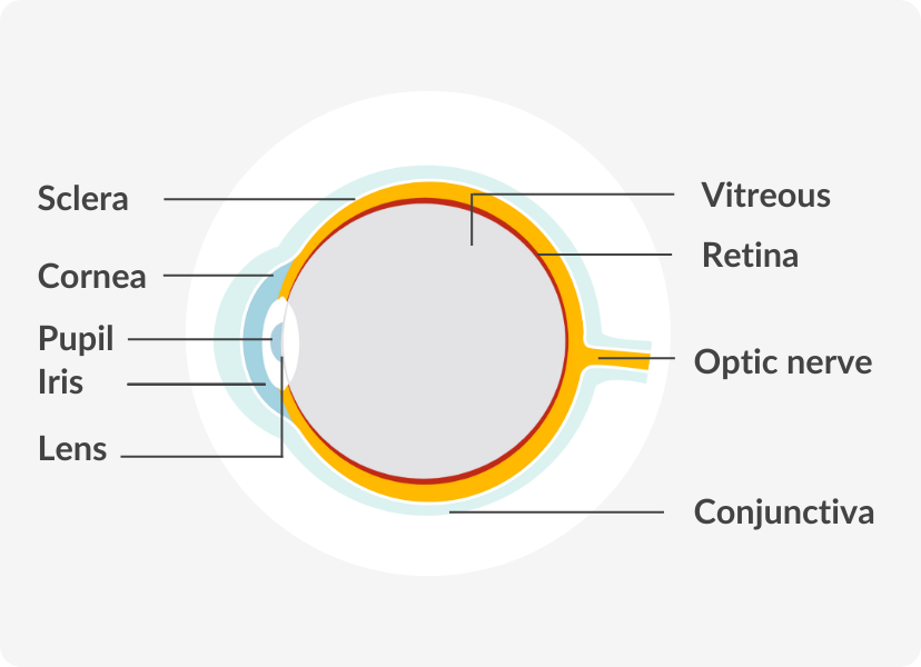
Eyeballs are shaped like spheres and sit in a protective socket called the orbit. The eyeball consists of three layers that surround a clear jelly. This jelly, known as the vitreous, is inside the eye and helps maintain its shape.
The outer layer of the eye consists of the sclera and the cornea. The iris, pupil and crystalline lens (lens) are in the middle layer. And the innermost layer of the human eye is called the retina. Here is a quick breakdown of those main structures:
- Cornea – the transparent structure at the front of the eye
- Sclera – the white part of the eye that surrounds the iris
- Iris – the coloured portion of the eye
- Pupil – the black circle in the center of the iris
- Lens – responsible for focusing light and images
- Retina – the light sensitive tissue at the back of the eye
How does the eye work?
The eye is attached to six muscles that control eye movement. One muscle moves the eye right, one to the left and the other four move the eye up, down and on an angle.
Human eyes can see about 200 degrees in all directions, including in front of them and to the sides (AAO, 2023). The anatomy of the eye is complex, allowing us to see images, movement, depth and millions of colours.
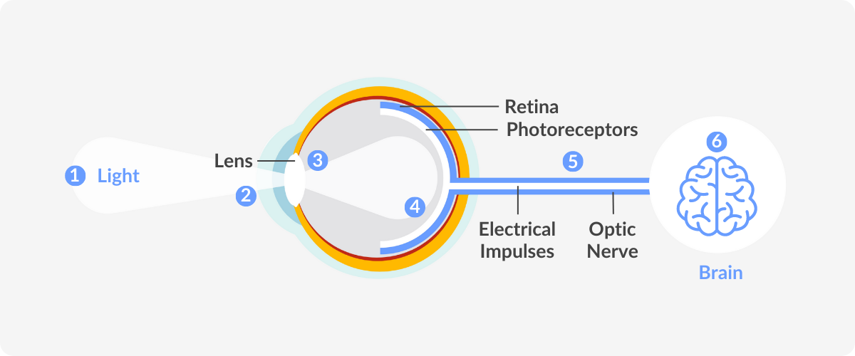
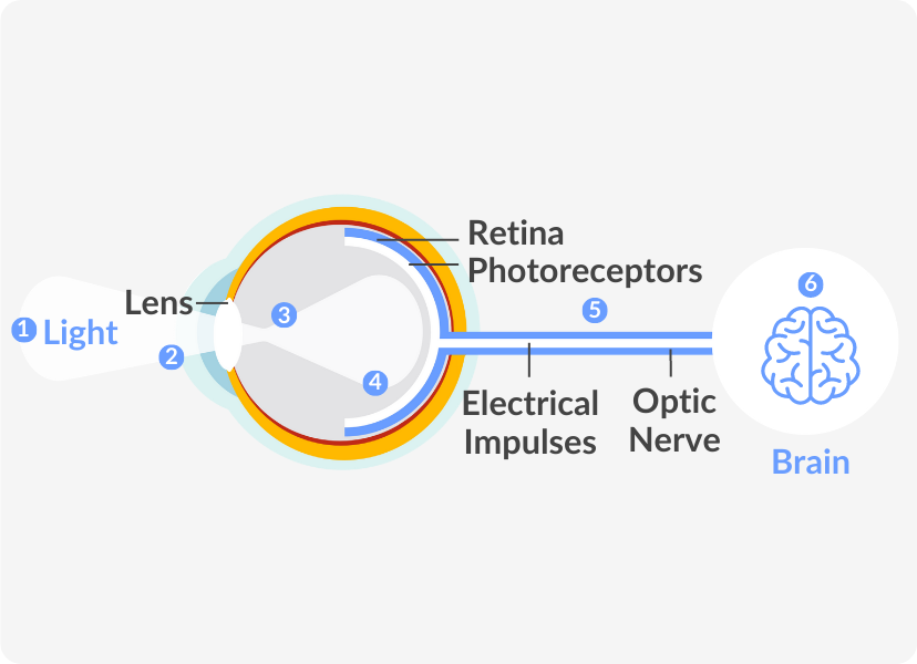
The different parts of the eye work together to send visual information to the brain. When we see an object, we are actually seeing light reflecting off that object.
Sight starts at the cornea and ends in the brain. Light passes through the cornea to the lens. Here, light is focused on the retina. When light reaches the retina, at the back of the eye, images are changed into signals and sent through the optic nerve to the brain, allowing us to comprehend these images.
Each component of the surface, the middle and the inner layers has a role to play in the whole process, all working together, be it directly or indirectly.
The eye is attached to six muscles that control eye movement. One muscle moves the eye right, one to the left and the other four move the eye up, down and on an angle.
Human eyes can see about 200 degrees in all directions, including in front of them and to the sides (AAO, 2023). The anatomy of the eye is complex, allowing us to see images, movement, depth and millions of colours.
The different parts of the eye work together to send visual information to the brain. When we see an object, we are actually seeing light reflecting off that object.
Sight starts at the cornea and ends in the brain. Light passes through the cornea to the lens. Here, light is focused on the retina.
When light reaches the retina, at the back of the eye, images are changed into signals and sent through the optic nerve to the brain, allowing us to comprehend these images.
Each component of the surface, the middle and the inner layers has a role to play in the whole process, all working together, be it directly or indirectly.
The surface of the eye
The surface of the eye is covered with a clear membrane called the conjunctiva and is protected by the eyelids. Our eyelids are folds of tissue that are also lined with conjunctiva.
Eyelids are vital in keeping the cornea moist and protecting the eyes from foreign objects and bright light or excess light. During waking hours, they lay mucous over the eyes, and during sleep prevent evaporation of the mucous.
Behind the cornea is the anterior chamber. The anterior chamber is filled with a fluid called aqueous humour.
Aqueous humour is constantly produced and drained from the eye to maintain normal eye pressure. The aqueous humour also provides nutrients to the lens and cornea, which do not have blood vessels (AAO, 2023).
DID YOU KNOW?
Our eyes have over two million moving parts and contain our body’s most active muscles (CAO, 2023).
The middle layer of the eye
The iris and pupil are located behind the anterior chamber in the central portion of the eye. They work together to control the amount of light that reaches the lens. The lens is directly behind the pupil and its main function is to focus light towards the back of the eye.
Similar to a camera, the eye can adjust the amount of light that enters so that it can function in different lighting conditions, from dim to very bright light.
The aperture control on your camera will let more or less light in depending on the surrounding environment. In our eyes, muscles in the iris dilate (widen) or constrict (narrow) the pupil to control how much light enters the lens.
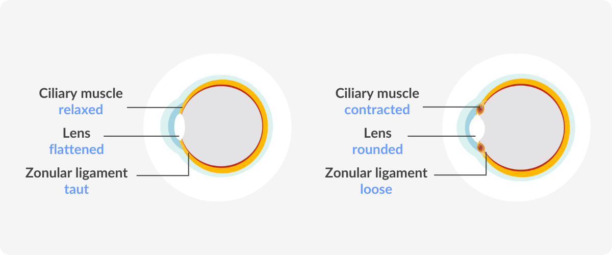
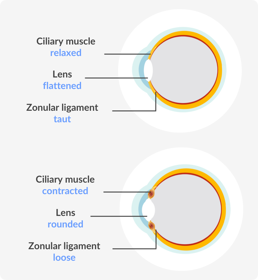
The ciliary body is located behind the iris and has small muscles that are attached to the lens. Together with the zonules, tiny thread-like fibres that hold the eye’s lens in place, these muscles allow the lens to change shape and focus on near or far objects.
When our eyes look at something very close to us, the zonules loosen and the lens thickens. When the eye looks at images far away, the muscles relax, the zonules tighten and the lens decreases in thickness. This causes images far away to come into focus.
Unlike a camera lens, which moves closer or further from an object to focus, the human lens does not move position but changes shape. When looking at a distant object, the lens becomes flattened. On the other hand, when looking at an object up close, the lens becomes rounded.
The iris and pupil are located behind the anterior chamber in the central portion of the eye. They work together to control the amount of light that reaches the lens.
The lens is directly behind the pupil and its main function is to focus light towards the back of the eye.
Similar to a camera, the eye can adjust the amount of light that enters so that it can function in different lighting conditions, from dim to very bright light.
The aperture control on your camera will let more or less light in depending on the surrounding environment. In our eyes, muscles in the iris dilate (widen) or constrict (narrow) the pupil to control how much light enters the lens.
The ciliary body is located behind the iris and has small muscles that are attached to the lens. Together with the zonules, tiny thread-like fibres that hold the eye’s lens in place, these muscles allow the lens to change shape and focus on near or far objects.
When our eyes look at something very close to us, the zonules loosen and the lens thickens. When the eye looks at images far away, the muscles relax, the zonules tighten and the lens decreases in thickness. This causes images far away to come into focus.
Unlike a camera lens, which moves closer or further from an object to focus, the human lens does not move position but changes shape.
When looking at a distant object, the lens becomes flattened. On the other hand, when looking at an object up close, the lens becomes rounded.
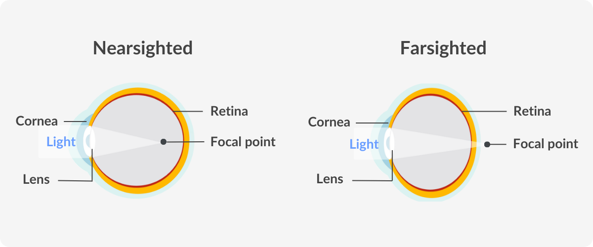
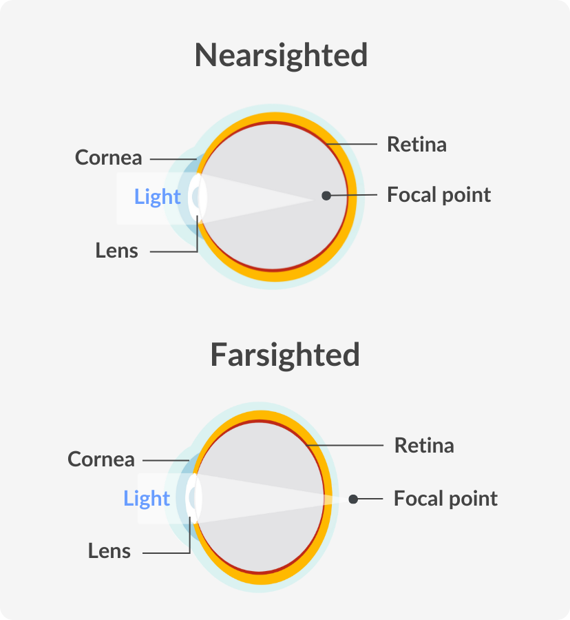
After the lens focuses incoming light rays it reflects it on the retina. Refractive errors like farsightedness or nearsightedness occur when the lens improperly focuses images on the retina.
If the lens focuses light behind the retina, this leads to farsightedness. If the lens focuses light in front of the retina, nearsightedness occurs. You must speak with an eye doctor should you notice any differences in your vision. Prescription glasses or contact lenses may help in correcting these issues.
After the lens focuses incoming light rays it reflects it on the retina. Refractive errors like farsightedness or nearsightedness occur when the lens improperly focuses images on the retina.
If the lens focuses light behind the retina, this leads to farsightedness. If the lens focuses light in front of the retina, nearsightedness occurs.
You must speak with an eye doctor should you notice any differences in your vision. Prescription glasses or contact lenses may help in correcting these issues.
The inner layer of the eye
Once the retina senses the light from the lens, it is responsible for converting it into electrical signals to send to the brain. The retina comprises two parts, the macula, and the peripheral retina.
In the centre, the macula processes most of what is directly in view, or your central vision. The peripheral retina is responsible for your peripheral vision.
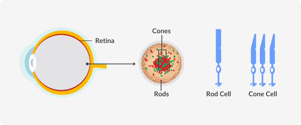
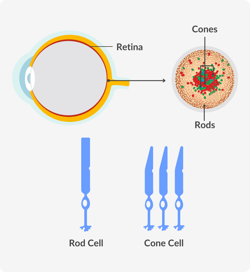
The retina is made up of many different light-sensitive cells. Photoreceptors are a type of cell that turn light into an electric signal so the brain can compute images.
Rods and cones are photoreceptors that work together to give a clear picture of what you’re seeing. Rods are sensitive to light and allow us to see in low-light conditions. Cones perceive colour and fine details and provide a sharp, accurate image (AAO, 2023).
These electrical impulses are sent through the optic nerve to the visual cortex, the part of the brain responsible for sight. The optic nerve is made up of millions of nerve fibres and is essential in transmitting all visual information, including brightness, perception, colour and contrast (AAO, 2023).
Once the retina senses the light from the lens, it is responsible for converting it into electrical signals to send to the brain. The retina comprises two parts, the macula, and the peripheral retina.
In the centre, the macula processes most of what is directly in view, or your central vision. The peripheral retina is responsible for your peripheral vision.
The retina is made up of many different light-sensitive cells. Photoreceptors are a type of cell that turn light into an electric signal so the brain can compute images.
Rods and cones are photoreceptors that work together to give a clear picture of what you’re seeing.
Rods are sensitive to light and allow us to see in low-light conditions. Cones perceive colour and fine details and provide a sharp, accurate image (AAO, 2023).
These electrical impulses are sent through the optic nerve to the visual cortex, the part of the brain responsible for sight.
The optic nerve is made up of millions of nerve fibres and is essential in transmitting all visual information, including brightness, perception, colour and contrast (AAO, 2023).
What conditions affect how the eyes work?
Various conditions can affect how our eyes function. Common vision problems like nearsightedness, farsightedness and astigmatism, as well as eye injuries, are among these issues.
Problems with the muscles in the eyes are common. Strabismus (crossed eyes) or amblyopia (lazy eye) can lead to physical and visual changes in the eye.
As we age, our eyes may change. Many people begin to lose close-range vision around the age of 45 (Cleveland Clinic, 2021) and may develop presbyopia. The lens may start to deteriorate and lead to the need for reading glasses. In more severe cases, a cloudy lens may be replaced with an intraocular lens through surgery.
Common problems like infection or irritation can cause redness, swelling or discomfort. If you notice changes in your vision or eyes, contact your eye doctor for a check-up.
Various conditions can affect how our eyes function. Common vision problems like nearsightedness, farsightedness and astigmatism, as well as eye injuries, are among these issues.
Problems with the muscles in the eyes are common. Strabismus (crossed eyes) or amblyopia (lazy eye) can lead to physical and visual changes in the eye.
As we age, our eyes may change. Many people begin to lose close-range vision around the age of 45 (Cleveland Clinic, 2021) and may develop presbyopia.
The lens may start to deteriorate and lead to the need for reading glasses. In more severe cases, a cloudy lens may be replaced with an intraocular lens through surgery.
Common problems like infection or irritation can cause redness, swelling or discomfort. If you notice changes in your vision or eyes, contact your eye doctor for a check-up.
How can I keep my eyes healthy?
Eye anatomy is complex, and we must take care of our eyes in order for everything to function correctly. It is crucial to see an eye doctor regularly for eye exams to maintain eye health. If required and prescribed, wearing corrective lenses is vital to prevent further damage.
If you spend a lot of time in front of digital screens, consider wearing computer glasses or taking breaks and doing eye exercises. When outside, sunglasses with proper UV protection are essential.
Always wear protective eyewear during contact sports or if you have a job working with tools that could potentially lead to eye injuries.
If you have any questions about maintaining eye health, head to our Optical Centre and speak to one of our certified Opticians.
Reference List
https://www.aao.org/eye-health/anatomy/parts-of-eye
Canadian Association of Optometrists. (2023, April 12). Fun Eye Facts. Https://Opto.ca/. Retrieved July 10, 2023, from
https://opto.ca/eye-health-library/fun-eye-facts
Cleveland Clinic (2021, September 20). Eyes. Https://my.Clevelandclinic.org/. Retrieved July 10, 2023, from
https://my.clevelandclinic.org/health/body/21823-eyes