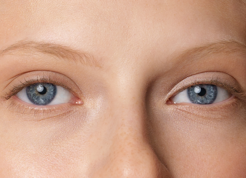
Tunnel Vision

By Anna MacGabhann
Reviewed by Beck Jinnette
Beck Jinnette

Reviewed by

Beck Jinnette
Beck has over 17 years of experience in eye care, holding her Certificate IV in Dispensing in Australia.
Do you ever feel like your world is slowly shrinking? You’re not alone. Tunnel vision is something that many people suffer from for various reasons.
Tunnel Vision example

Disclaimer:  We at SmartBuyGlasses are not medical doctors. This article contains general advice. If you experience tunnel vision, please consult your doctor or an eye care professional for treatment.

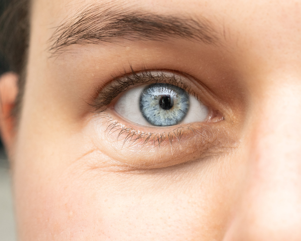
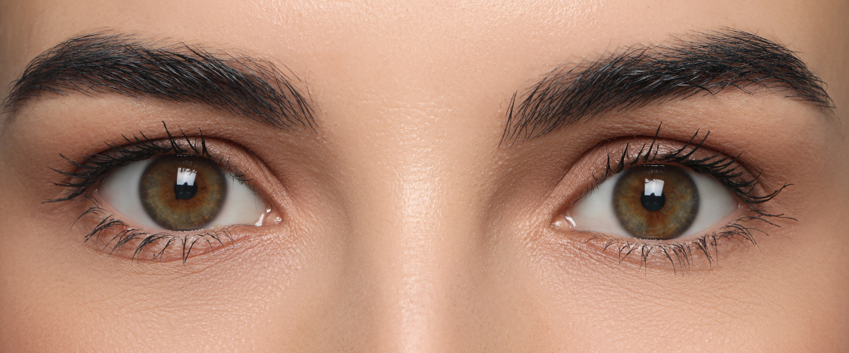
What is tunnel vision or PVL?

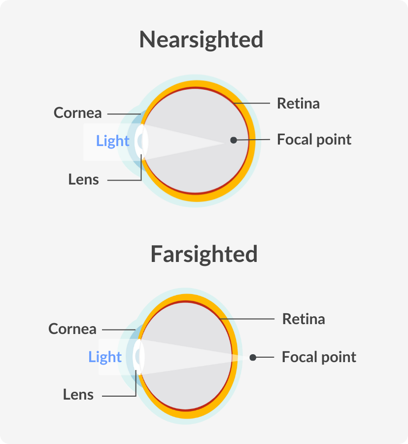
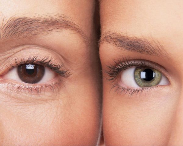
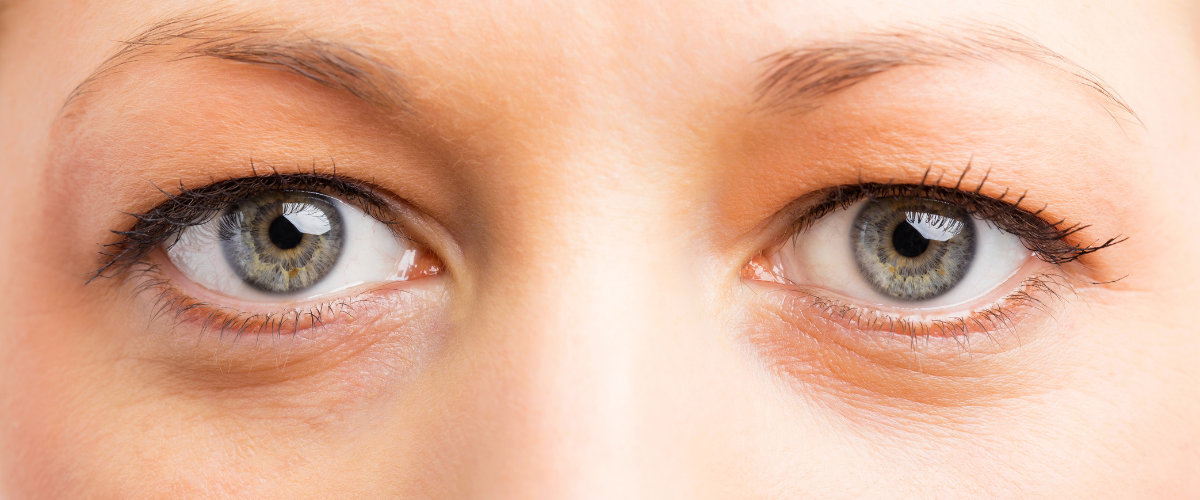
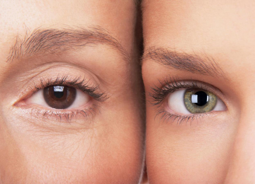
Tunnel vision, also known as Peripheral Vision Loss (PVL), is a medical condition where an individual experiences a narrowed field of vision. This condition can occur due to various reasons, such as optic nerve damage or brain injuries. 

When tunnel vision occurs, people may find it difficult to see objects on the peripheries of their field of vision and might have trouble with activities that require them to scan their surroundings.

While for some individuals, tunnel vision develops over time, others may experience it suddenly due to an underlying medical condition. Although it’s not always treatable, there are various aids and treatments available to help people with PVL cope with the changes in their vision.

What is an example of tunnel vision?

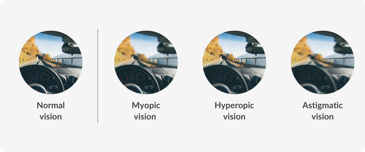
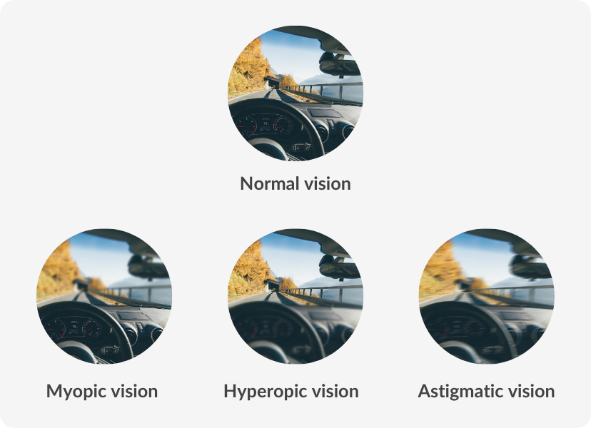
Tunnel vision is a term used to describe a type of visual impairment where the individual’s side vision is severely reduced, resulting in a narrow visual field. This can lead to a person feeling as though they are looking through a tunnel, with everything outside the tunnel being blurry or completely out of sight.

What causes tunnel vision?

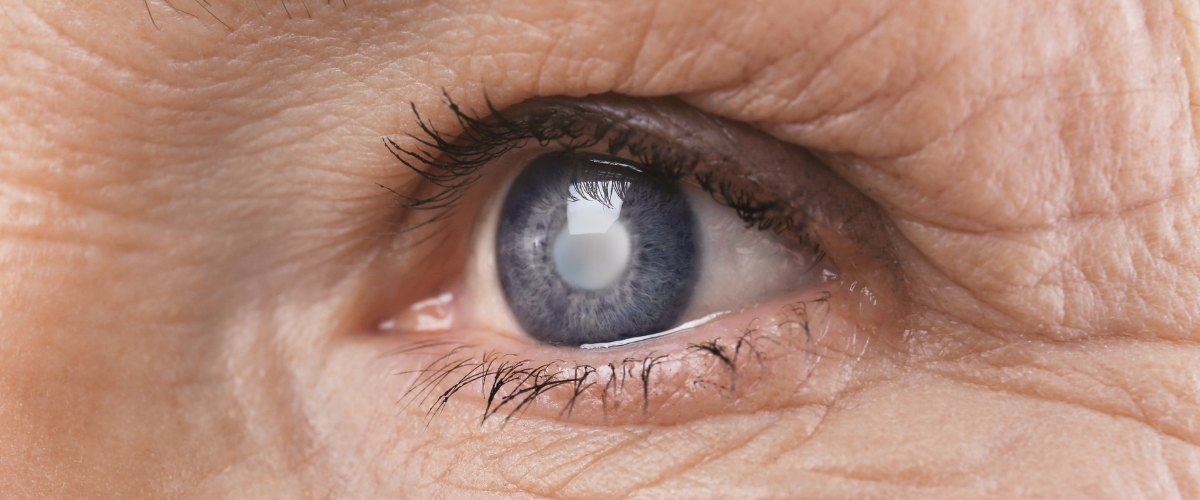
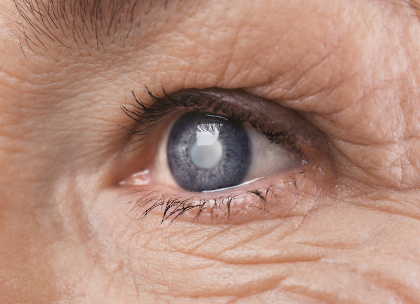
The causes of tunnel vision or tubular vision can vary from person to person and can be brought about by both physical and psychological factors. Physically, it can be caused by eye diseases such as glaucoma, cataracts or retinitis pigmentosa, among others. 

On the other hand, psychological causes such as panic attacks can also trigger tunnel vision as a way of the body protecting itself from a perceived threat.
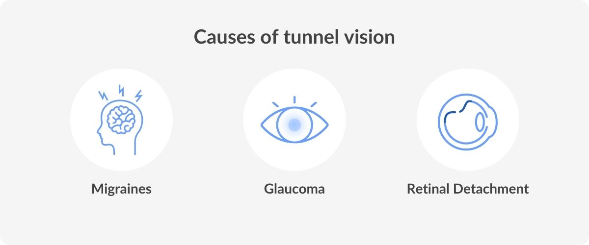
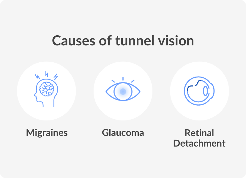
Migraine

Migraines can cause a range of visual disturbances, including tunnel vision, compromised vision on one side or total vision loss. Migraines can result from various ocular and nervous stimuli and can last anywhere from 10 minutes to multiple hours.

Retinal detachment

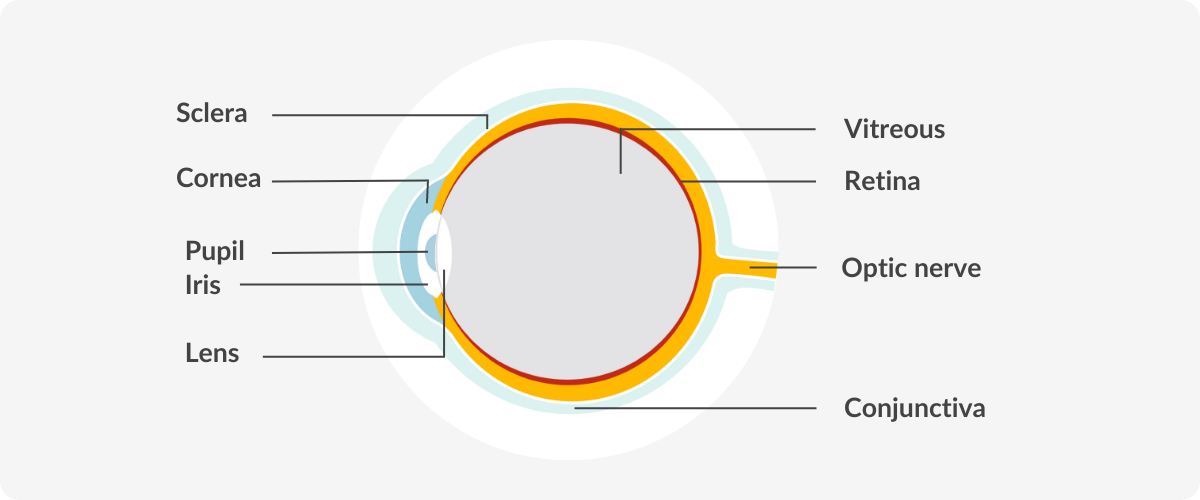
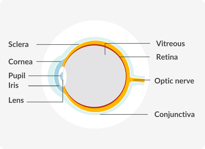
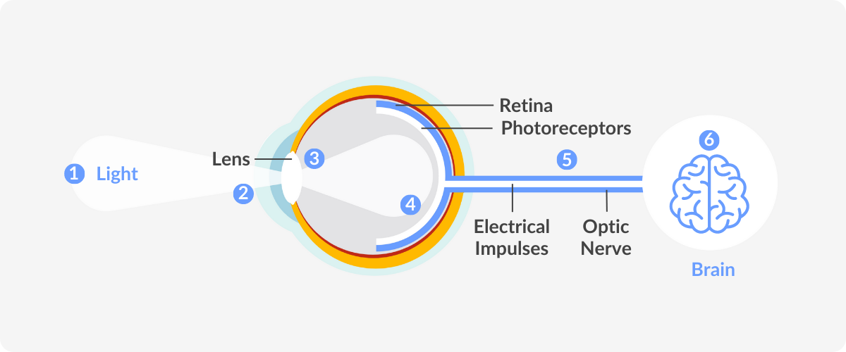
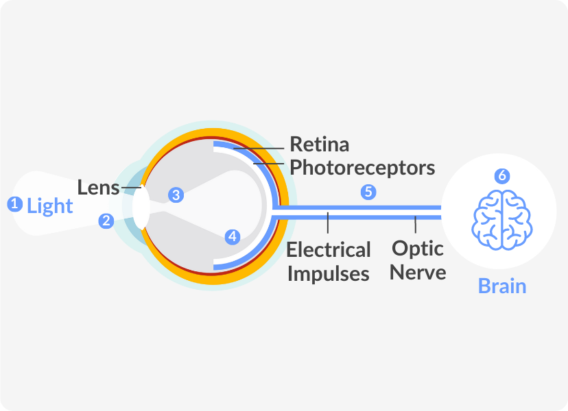
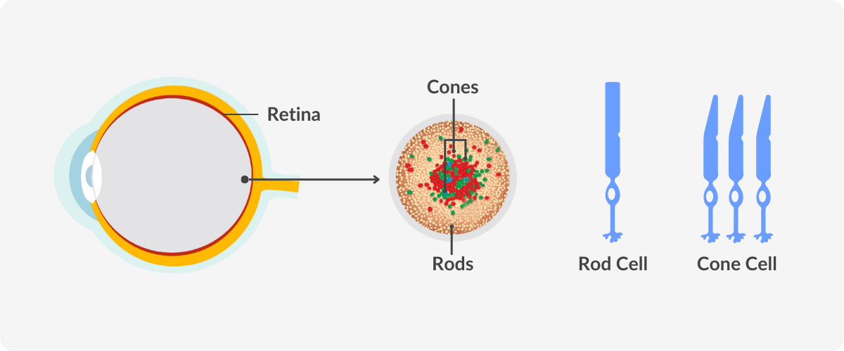
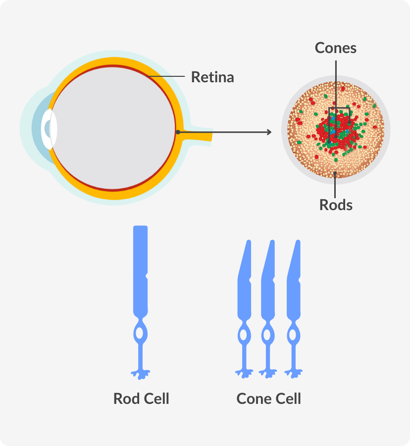
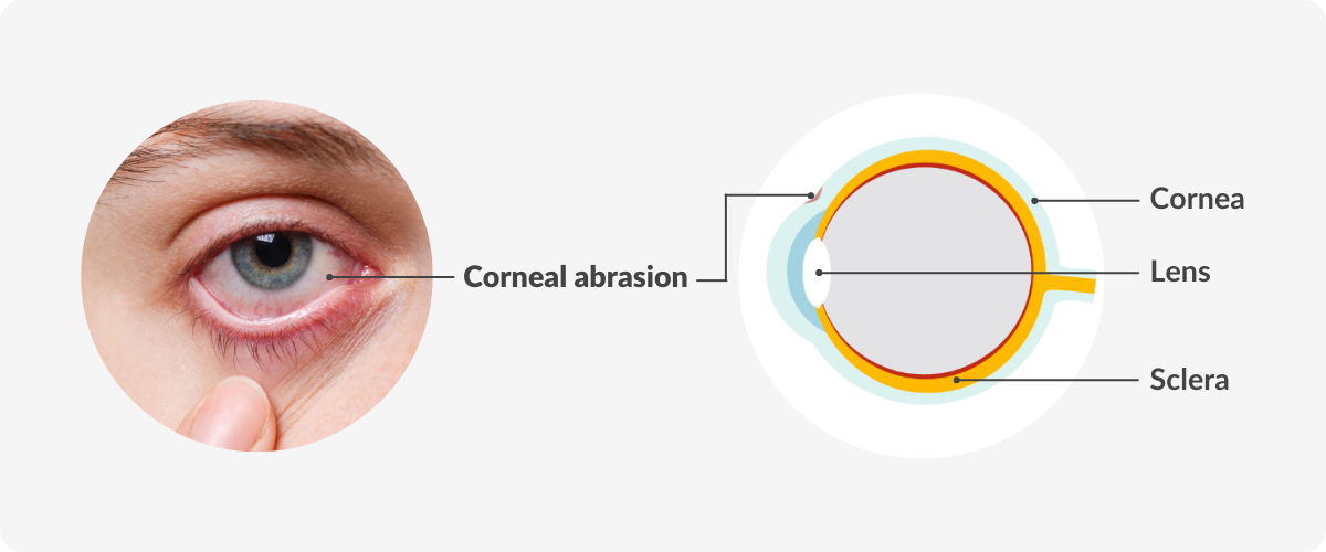
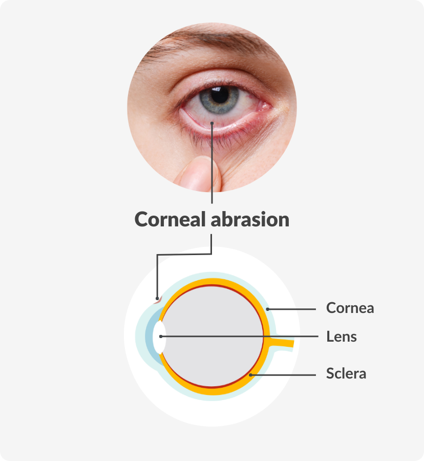
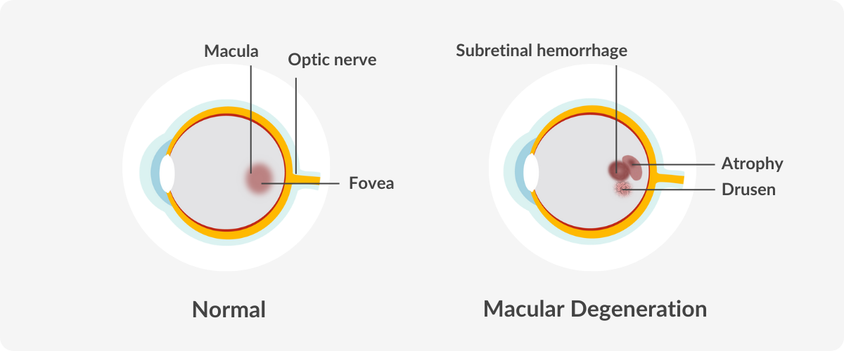
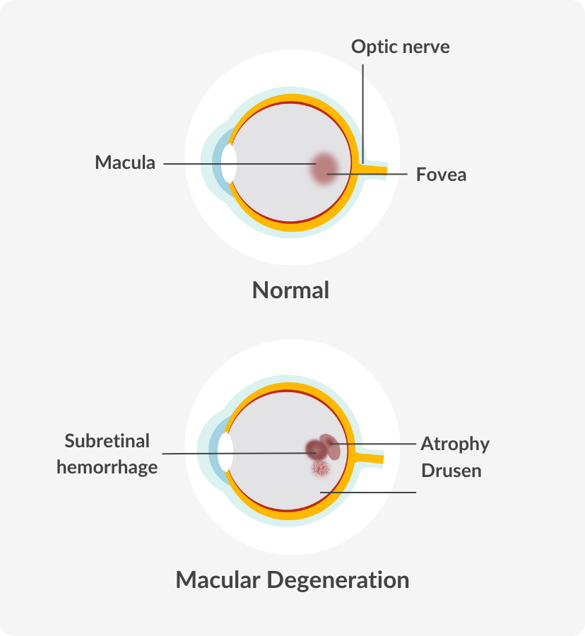
Retinal detachment occurs when the retina, located at the back of the eye, separates from the underlying tissue. The optic nerve, responsible for transmitting visual information to the brain, is directly impacted by retinal detachment, resulting in tunnel vision. Any changes in vision should be immediately evaluated by an eye care professional through a comprehensive dilated eye exam.

Glaucoma

Glaucoma is a medical condition that affects the eyes. It is caused by a buildup of pressure from excess fluid within the eye, which can result in peripheral vision loss. Glaucoma can lead to loss of vision if it’s not diagnosed and treated early.

Stroke

A stroke is a medical condition that occurs when the blood supply to part of the brain is reduced. It can happen when blood vessels in the brain are blocked. One of the first symptoms of a stroke is loss to peripheral vision, resulting in a concentration on the  central visual field.

causes of tunnel vision

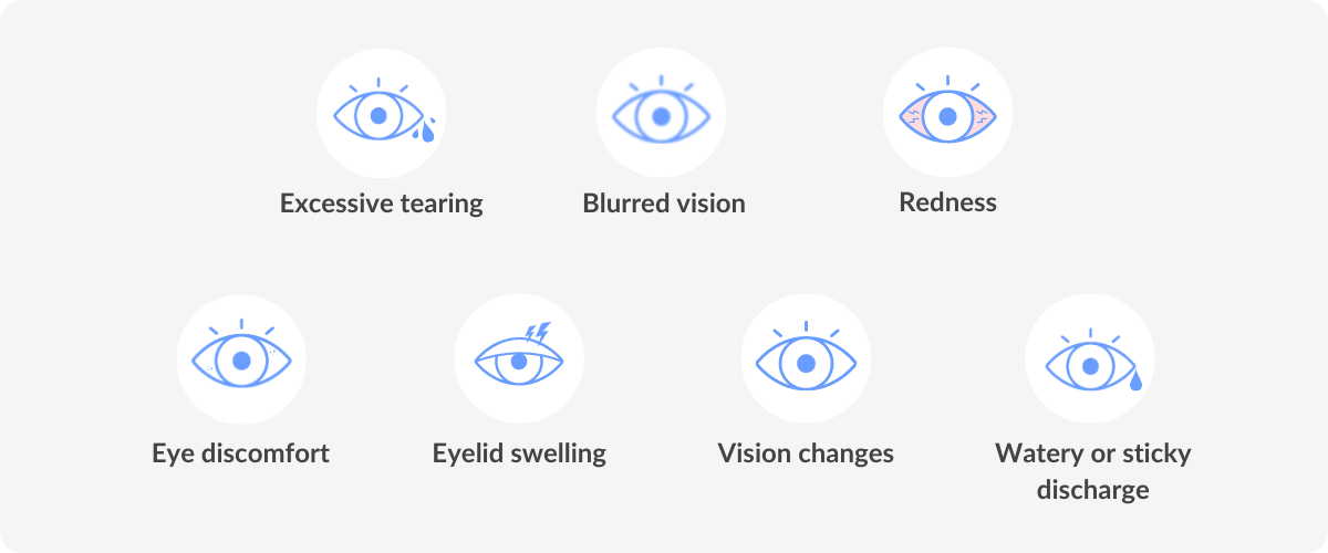
Symptoms of tunnel vision

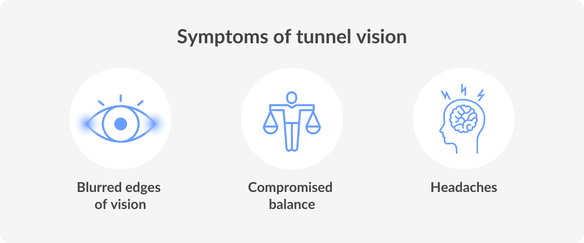
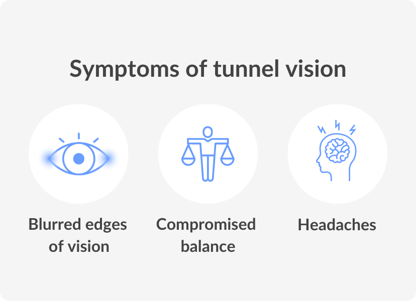
Tunnel vision affects an individual’s peripheral vision, causing them to see only a narrow channel while the rest of the surroundings become blurred or invisible. The symptoms are usually gradual and can go unnoticed at first. 

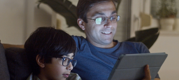
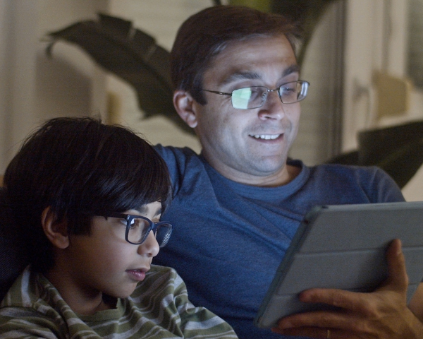
If you start experiencing visual disturbances such as difficulty focusing on objects that are outside of your central vision, altered night vision or are having a hard time adjusting to dim lights, you may be experiencing tunnel vision symptoms.

Other symptoms include eye fatigue and headaches, especially when trying to focus for an extended period. In extreme cases, tunnel vision can cause a complete loss of peripheral vision, resulting in difficulty with balance. It is vital to consult a specialist or eye doctor if you believe you may be experiencing tunnel vision symptoms.

symptoms of tunnel vision

Is tunnel vision a symptom of anxiety?

When it comes to anxiety, there are a multitude of symptoms that can manifest themselves in different ways. One of these symptoms that people may experience is tunnel vision. It is important to remember that it is a common symptom of anxiety and can be managed with the right tools and techniques.

These could include things like deep breathing exercises, meditation or talking to a mental health professional to work through the root causes of your anxiety. Overall, it is important to remember that tunnel vision is just one symptom of anxiety and with the right support and resources, you can learn to manage and overcome it.

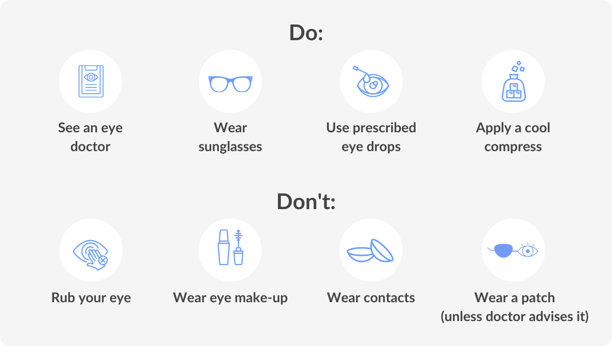
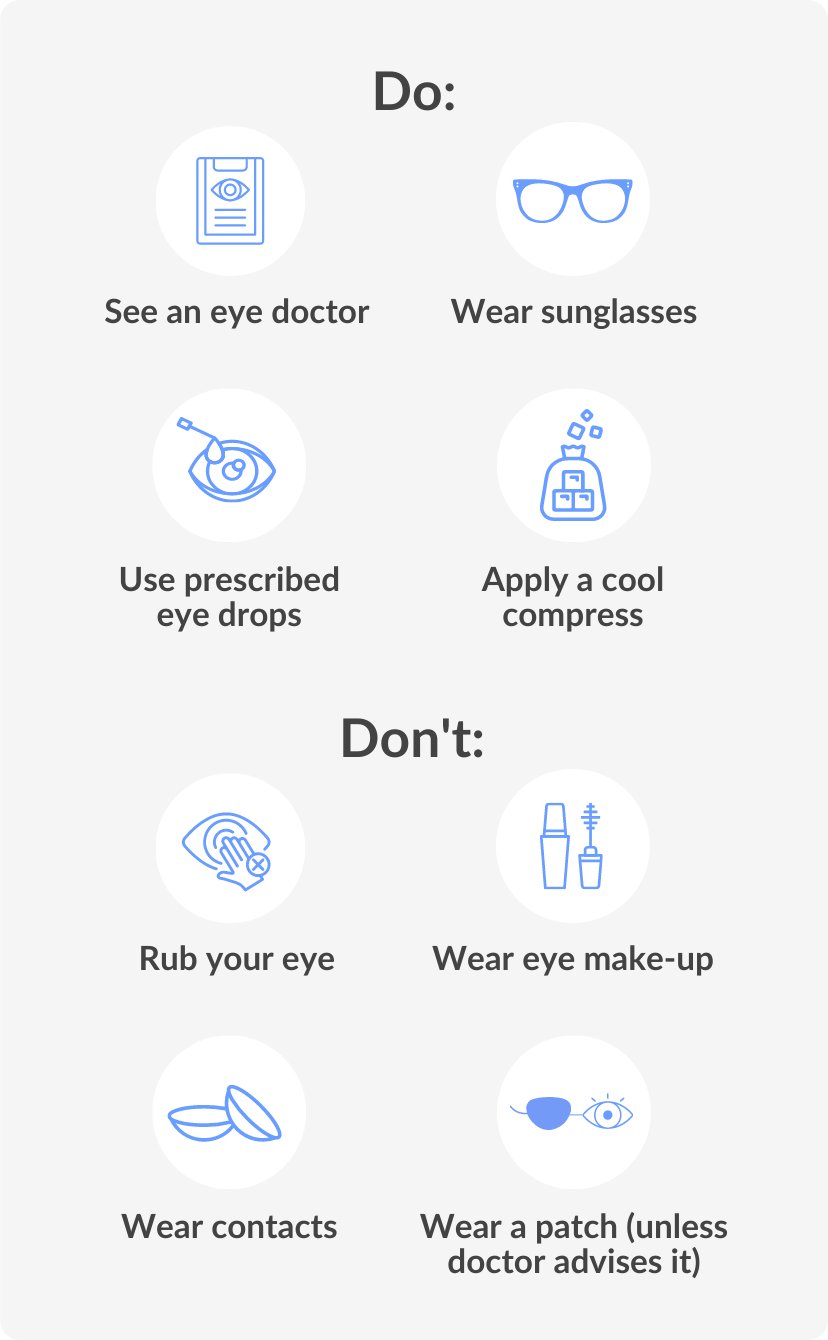
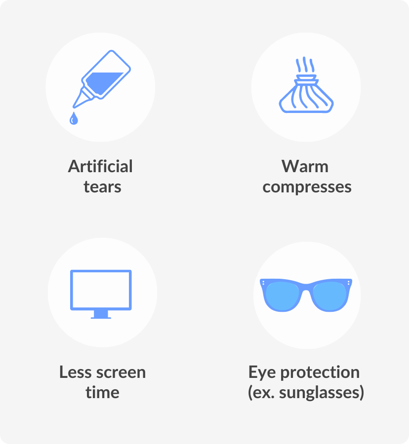
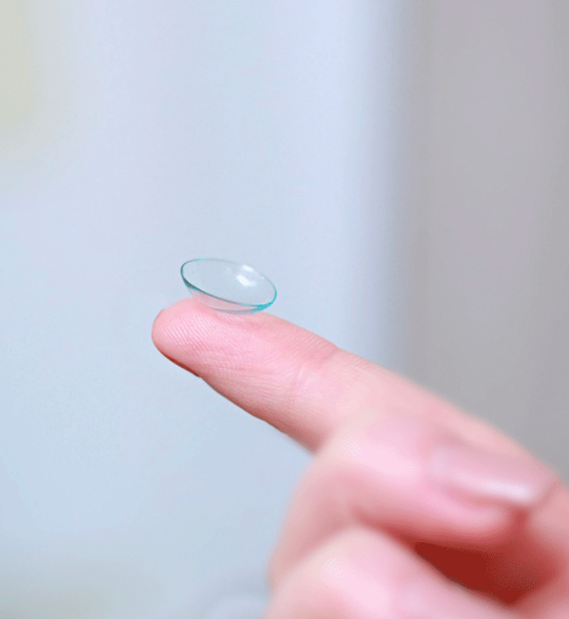
How do you treat tunnel vision?

We recommend that you consult your eyecare professional to discuss the treatment of tunnel vision as there are many factors to consider. Overall, when your doctor decides a course of treatment for tunnel vision, they will address the underlying cause. 

Depending on the cause, they may recommend medications, surgery or lifestyle changes. For example, if your tunnel vision is caused by high blood pressure, you may be prescribed medication to control your blood pressure.

If it is caused by a migraine, your doctor may recommend migraine medication or lifestyle changes such as reducing stress, eating a healthy diet and getting regular exercise. Whatever the cause may be, it is vital to seek medical attention. This can be a serious condition so your doctor will take a course of action to suit the severity of your case.

DID YOU KNOW?

Tunnel vision is caused by a number of health problems. Addressing the cause of this visual disturbance as soon as possible will help you make a faster recovery.

Is PVL or tunnel vision permanent?

The good news is PVL does not usually result in permanent vision loss. It’s a transient condition that occurs due to a variety of causes. It’s not uncommon to experience PVL or tunnel vision during high-pressure situations like exams or job interviews. 

However, it’s important to seek medical help if you experience PVL or tunnel vision frequently, as it could be a symptom of an underlying medical condition. Remember, taking care of your mental and physical health is extremely important and seeking professional help when needed is nothing to be ashamed of.

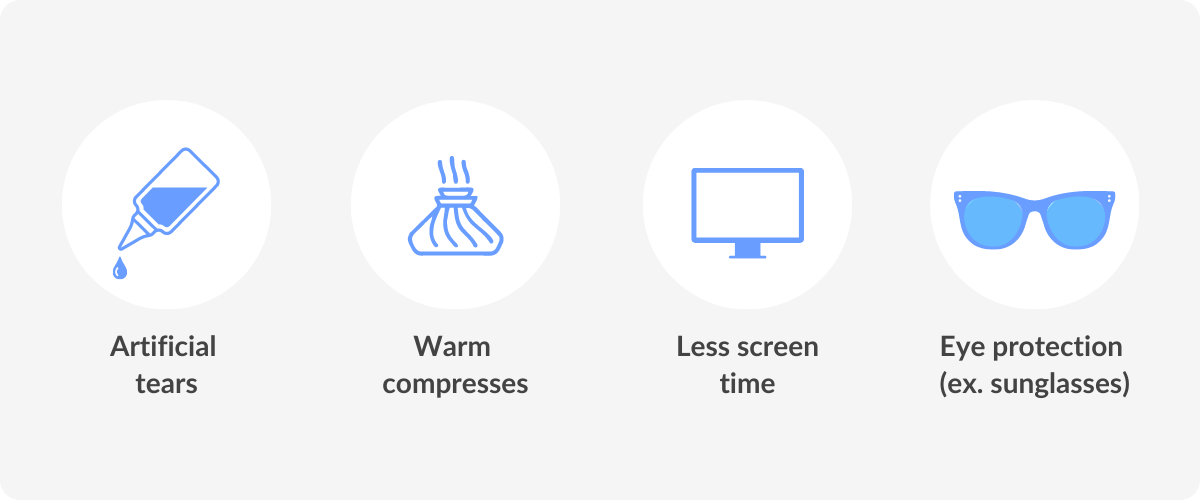
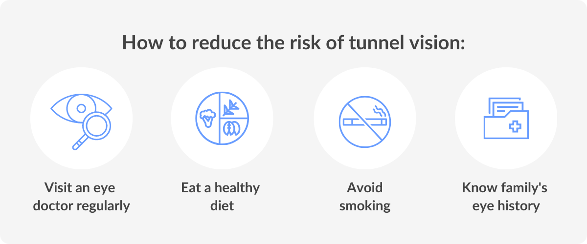
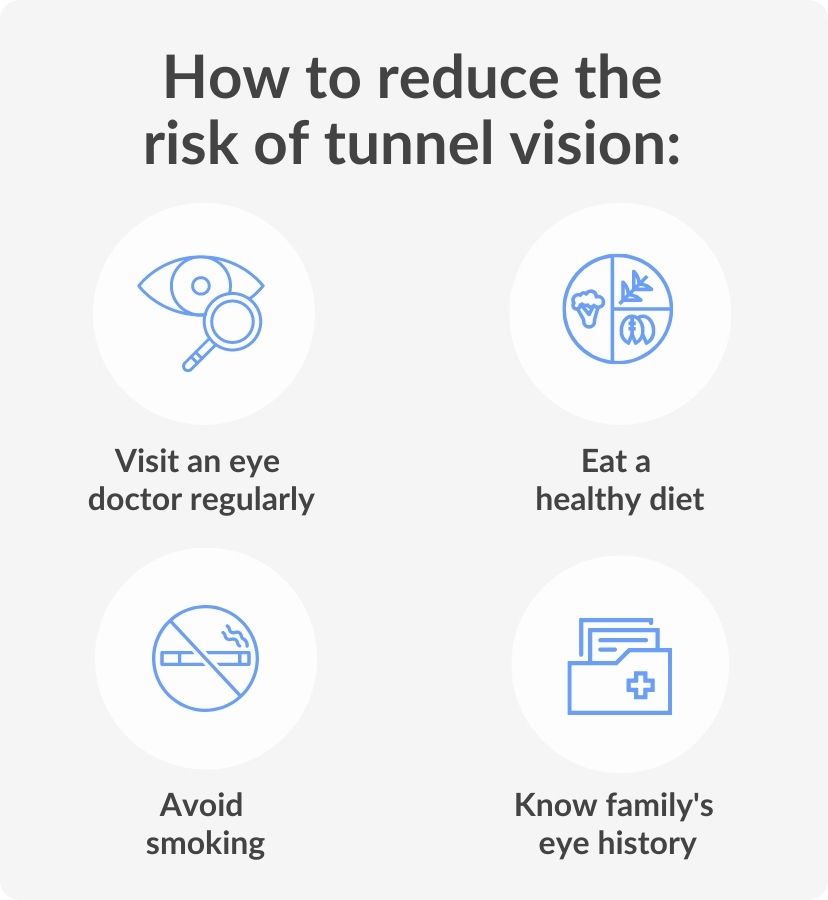
How to reduce the risk of tunnel vision

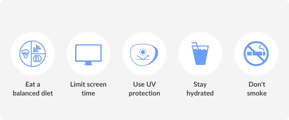
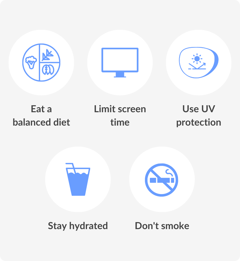
Our eyesight is precious and it’s important to take steps to protect it. One potential threat to healthy vision is tunnel vision. Fortunately, there are steps you can take to reduce the risk of developing tunnel vision.

Firstly, make sure you know your family’s eye health history and visit the eye doctor regularly. Secondly, ensure that your workspace is well-lit, as dimly lit environments can cause undue strain on your eyes. 

Finally, consider a well-balanced and nutritious diet with plenty of eye-healthy vitamins and make lifestyle choices to optimise personal health, such as avoiding smoking.

reducing the risk
reducing the risk of tunnel vision

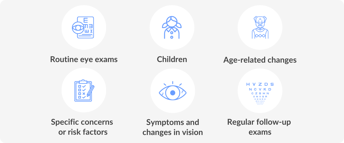
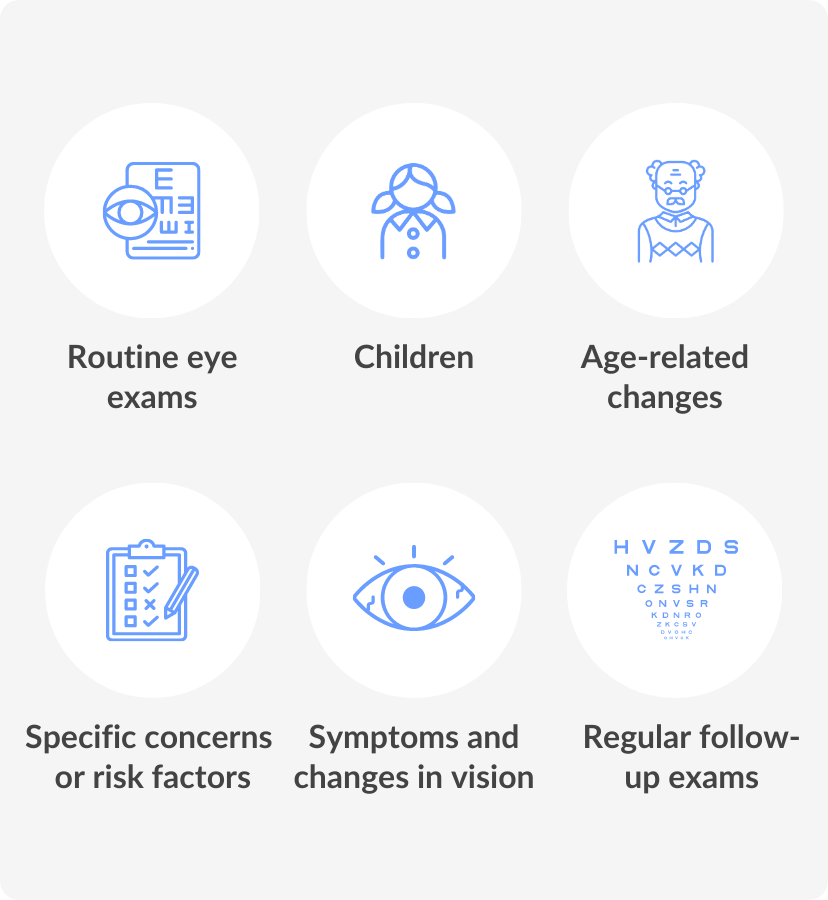
When to see a professional

Seeking help early on can often make the biggest difference in long-term outcomes. If you’re experiencing persistent symptoms or concerns that are affecting your daily life, it’s worth reaching out to a professional as soon as possible for support. 

They can help you better understand your condition and provide appropriate treatments or referrals to specialists if necessary. At SmartBuyGlasses Optical Centre, we are here to reassure you with the facts so you can make informed choices about your health before visiting a doctor.

Eye Anatomy

By Claire Capuano
Reviewed by Beck Jinnette
Beck Jinnette

Reviewed by

Beck Jinnette
Beck has over 17 years of experience in eye care, holding her Certificate IV in Dispensing in Australia.
Our eyes are made up of many parts that work together with the brain. They are critical in helping us interact with the world.
image of a persons eye
SUMMARY

The eye is a complex organ of the body.

The key parts include the cornea, lens, retina and optic nerve; each plays a role in capturing and processing light.

By understanding how the eye works, you can better recognise symptoms of vision problems and maintain eye health.

Human beings have five senses, sight being one of them. Eyes are responsible for giving us the sense of vision and allow us to see and experience all the beauty that surrounds us. 

One of the most important organs in the body, eyes play a major role in day-to-day life and experiences. “Eyes are the windows to the soul” is an expression that is often used to describe the deep connection we feel when making eye contact with someone. 

It is through our eyes and the sense of sight that we are able to connect with people, places and things. Small but mighty, it is essential to keep our eyes healthy. Most people tend to take sight for granted, without giving much thought to how it is all made possible. 

Human beings have five senses, sight being one of them. Eyes are responsible for giving us the sense of vision and allow us to see and experience all the beauty that surrounds us. 

One of the most important organs in the body, eyes play a major role in day-to-day life and experiences. 

“Eyes are the windows to the soul” is an expression that is often used to describe the deep connection we feel when making eye contact with someone. It is through our eyes and the sense of sight that we are able to connect with people, places and things. 

Small but mighty, it is essential to keep our eyes healthy. Most people tend to take sight for granted, without giving much thought to how it is all made possible.

What makes up an eye?

The human eye is a complex organ that enables us to perceive the world around us by capturing and processing light. The eye is made up of many light-sensitive cells and works in conjunction with the brain to provide us with the sense of vision. 

Eyeballs are shaped like spheres and sit in a protective socket called the orbit. The eyeball consists of three layers that surround a clear jelly. This jelly, known as the vitreous, is inside the eye and helps maintain its shape. 

The outer layer of the eye consists of the sclera and the cornea. The iris, pupil and crystalline lens (lens) are in the middle layer. And the innermost layer of the human eye is called the retina. Here is a quick breakdown of those main structures:

  • Cornea – the transparent structure at the front of the eye
  • Sclera – the white part of the eye that surrounds the iris
  • Iris – the coloured portion of the eye
  • Pupil – the black circle in the center of the iris
  • Lens – responsible for focusing light and images
  • Retina – the light sensitive tissue at the back of the eye
infographic of the eye

How does the eye work?

The eye is attached to six muscles that control eye movement. One muscle moves the eye right, one to the left and the other four move the eye up, down and on an angle.

Human eyes can see about 200 degrees in all directions, including in front of them and to the sides (AAO, 2023). The anatomy of the eye is complex, allowing us to see images, movement, depth and millions of colours.

The different parts of the eye work together to send visual information to the brain. When we see an object, we are actually seeing light reflecting off that object. 

Sight starts at the cornea and ends in the brain. Light passes through the cornea to the lens. Here, light is focused on the retina. When light reaches the retina, at the back of the eye, images are changed into signals and sent through the optic nerve to the brain, allowing us to comprehend these images.

Each component of the surface, the middle and the inner layers has a role to play in the whole process, all working together, be it directly or indirectly. 

The eye is attached to six muscles that control eye movement. One muscle moves the eye right, one to the left and the other four move the eye up, down and on an angle.

Human eyes can see about 200 degrees in all directions, including in front of them and to the sides (AAO, 2023). The anatomy of the eye is complex, allowing us to see images, movement, depth and millions of colours.

The different parts of the eye work together to send visual information to the brain. When we see an object, we are actually seeing light reflecting off that object. 

Sight starts at the cornea and ends in the brain. Light passes through the cornea to the lens. Here, light is focused on the retina. 

When light reaches the retina, at the back of the eye, images are changed into signals and sent through the optic nerve to the brain, allowing us to comprehend these images.

Each component of the surface, the middle and the inner layers has a role to play in the whole process, all working together, be it directly or indirectly. 

infographic of how we see

The surface of the eye

The surface of the eye is covered with a clear membrane called the conjunctiva and is protected by the eyelids. Our eyelids are folds of tissue that are also lined with conjunctiva. 

Eyelids are vital in keeping the cornea moist and protecting the eyes from foreign objects and bright light or excess light. During waking hours, they lay mucous over the eyes, and during sleep prevent evaporation of the mucous.

Behind the cornea is the anterior chamber. The anterior chamber is filled with a fluid called aqueous humour. 

Aqueous humour is constantly produced and drained from the eye to maintain normal eye pressure. The aqueous humour also provides nutrients to the lens and cornea, which do not have blood vessels (AAO, 2023).

DID YOU KNOW?

Our eyes have over two million moving parts and contain our body’s most active muscles (CAO, 2023).

The middle layer of the eye

The iris and pupil are located behind the anterior chamber in the central portion of the eye. They work together to control the amount of light that reaches the lens. The lens is directly behind the pupil and its main function is to focus light towards the back of the eye. 

Similar to a camera, the eye can adjust the amount of light that enters so that it can function in different lighting conditions, from dim to very bright light. 

The aperture control on your camera will let more or less light in depending on the surrounding environment. In our eyes, muscles in the iris dilate (widen) or constrict (narrow) the pupil to control how much light enters the lens. 

The ciliary body is located behind the iris and has small muscles that are attached to the lens. Together with the zonules, tiny thread-like fibres that hold the eye’s lens in place, these muscles allow the lens to change shape and focus on near or far objects.

When our eyes look at something very close to us, the zonules loosen and the lens thickens. When the eye looks at images far away, the muscles relax, the zonules tighten and the lens decreases in thickness. This causes images far away to come into focus.

Unlike a camera lens, which moves closer or further from an object to focus, the human lens does not move position but changes shape. When looking at a distant object, the lens becomes flattened. On the other hand, when looking at an object up close, the lens becomes rounded. 

The iris and pupil are located behind the anterior chamber in the central portion of the eye. They work together to control the amount of light that reaches the lens. 

The lens is directly behind the pupil and its main function is to focus light towards the back of the eye. 

Similar to a camera, the eye can adjust the amount of light that enters so that it can function in different lighting conditions, from dim to very bright light. 

The aperture control on your camera will let more or less light in depending on the surrounding environment. In our eyes, muscles in the iris dilate (widen) or constrict (narrow) the pupil to control how much light enters the lens. 

The ciliary body is located behind the iris and has small muscles that are attached to the lens. Together with the zonules, tiny thread-like fibres that hold the eye’s lens in place, these muscles allow the lens to change shape and focus on near or far objects.

When our eyes look at something very close to us, the zonules loosen and the lens thickens. When the eye looks at images far away, the muscles relax, the zonules tighten and the lens decreases in thickness. This causes images far away to come into focus.

Unlike a camera lens, which moves closer or further from an object to focus, the human lens does not move position but changes shape. 

When looking at a distant object, the lens becomes flattened. On the other hand, when looking at an object up close, the lens becomes rounded.

infographic of the eye seeing things close and far

After the lens focuses incoming light rays it reflects it on the retina. Refractive errors like farsightedness or nearsightedness occur when the lens improperly focuses images on the retina. 

If the lens focuses light behind the retina, this leads to farsightedness. If the lens focuses light in front of the retina, nearsightedness occurs. You must speak with an eye doctor should you notice any differences in your vision. Prescription glasses or contact lenses may help in correcting these issues. 

After the lens focuses incoming light rays it reflects it on the retina. Refractive errors like farsightedness or nearsightedness occur when the lens improperly focuses images on the retina. 

If the lens focuses light behind the retina, this leads to farsightedness. If the lens focuses light in front of the retina, nearsightedness occurs. 

You must speak with an eye doctor should you notice any differences in your vision. Prescription glasses or contact lenses may help in correcting these issues.

infographic of nearsighted and farsighted

The inner layer of the eye

Once the retina senses the light from the lens, it is responsible for converting it into electrical signals to send to the brain. The retina comprises two parts, the macula, and the peripheral retina. 

In the centre, the macula processes most of what is directly in view, or your central vision. The peripheral retina is responsible for your peripheral vision. 

The retina is made up of many different light-sensitive cells. Photoreceptors are a type of cell that turn light into an electric signal so the brain can compute images. 

Rods and cones are photoreceptors that work together to give a clear picture of what you’re seeing. Rods are sensitive to light and allow us to see in low-light conditions. Cones perceive colour and fine details and provide a sharp, accurate image (AAO, 2023). 

These electrical impulses are sent through the optic nerve to the visual cortex, the part of the brain responsible for sight. The optic nerve is made up of millions of nerve fibres and is essential in transmitting all visual information, including brightness, perception, colour and contrast (AAO, 2023). 

Once the retina senses the light from the lens, it is responsible for converting it into electrical signals to send to the brain. The retina comprises two parts, the macula, and the peripheral retina. 

In the centre, the macula processes most of what is directly in view, or your central vision. The peripheral retina is responsible for your peripheral vision. 

The retina is made up of many different light-sensitive cells. Photoreceptors are a type of cell that turn light into an electric signal so the brain can compute images. 

Rods and cones are photoreceptors that work together to give a clear picture of what you’re seeing. 

Rods are sensitive to light and allow us to see in low-light conditions. Cones perceive colour and fine details and provide a sharp, accurate image (AAO, 2023). 

These electrical impulses are sent through the optic nerve to the visual cortex, the part of the brain responsible for sight. 

The optic nerve is made up of millions of nerve fibres and is essential in transmitting all visual information, including brightness, perception, colour and contrast (AAO, 2023).

infographic of the retina, rod and cone cells

What conditions affect how the eyes work?

Various conditions can affect how our eyes function. Common vision problems like nearsightedness, farsightedness and astigmatism, as well as eye injuries, are among these issues. 

Problems with the muscles in the eyes are common. Strabismus (crossed eyes) or amblyopia (lazy eye) can lead to physical and visual changes in the eye.

As we age, our eyes may change. Many people begin to lose close-range vision around the age of 45 (Cleveland Clinic, 2021) and may develop presbyopia. The lens may start to deteriorate and lead to the need for reading glasses. In more severe cases, a cloudy lens may be replaced with an intraocular lens through surgery. 

Common problems like infection or irritation can cause redness, swelling or discomfort. If you notice changes in your vision or eyes, contact your eye doctor for a check-up.

Various conditions can affect how our eyes function. Common vision problems like nearsightedness, farsightedness and astigmatism, as well as eye injuries, are among these issues. 

Problems with the muscles in the eyes are common. Strabismus (crossed eyes) or amblyopia (lazy eye) can lead to physical and visual changes in the eye.

As we age, our eyes may change. Many people begin to lose close-range vision around the age of 45 (Cleveland Clinic, 2021) and may develop presbyopia

The lens may start to deteriorate and lead to the need for reading glasses. In more severe cases, a cloudy lens may be replaced with an intraocular lens through surgery. 

Common problems like infection or irritation can cause redness, swelling or discomfort. If you notice changes in your vision or eyes, contact your eye doctor for a check-up.

How can I keep my eyes healthy?

Eye anatomy is complex, and we must take care of our eyes in order for everything to function correctly. It is crucial to see an eye doctor regularly for eye exams to maintain eye health. If required and prescribed, wearing corrective lenses is vital to prevent further damage.

If you spend a lot of time in front of digital screens, consider wearing computer glasses or taking breaks and doing eye exercises. When outside, sunglasses with proper UV protection are essential. 

Always wear protective eyewear during contact sports or if you have a job working with tools that could potentially lead to eye injuries.

If you have any questions about maintaining eye health, head to our Optical Centre and speak to one of our certified Opticians. 

Reference List

American Academy of Ophthalmology (2023, April 29). Eye Anatomy: Parts of the Eye and How We See. Https://www.aao.org/. Retrieved July 10, 2023, from
https://www.aao.org/eye-health/anatomy/parts-of-eye
Canadian Association of Optometrists. (2023, April 12). Fun Eye Facts. Https://Opto.ca/. Retrieved July 10, 2023, from
https://opto.ca/eye-health-library/fun-eye-facts
Cleveland Clinic (2021, September 20). Eyes. Https://my.Clevelandclinic.org/. Retrieved July 10, 2023, from
https://my.clevelandclinic.org/health/body/21823-eyes

What Does 20/20 Vision Mean?

Ask our dispensing opticians

Ask now
By Emma Moletto
Reviewed by Beck Jinnette
Beck Jinnette

Reviewed by

Beck Jinnette
Beck has over 17 years of experience in eye care, holding her Certificate IV in Dispensing in Australia.
Discover what 20/20 vision means and how it relates to normal eyesight.
vision eye test
SUMMARY

20/20 vision refers to normal visual acuity, meaning that you can see an object clearly from the standard distance for eye tests (20 feet away).

Since it only measures the clarity of distance vision, it doesn’t necessarily mean perfect eyesight, although people with 20/20 vision typically don’t require glasses.

Have you ever heard this term and wondered “what does 20/20 vision mean?” This article provides the explanation you’ve been looking for.

20/20 vision means that you can see an object clearly 20 feet (6m) away. It is a term used to indicate visual acuity (how clearly you can see).

For example, if you have 20/30 vision, that means that you can see at 30 feet (9m) what an average person with normal vision can see at 20 feet. In short, 20/20 vision is normal visual acuity.

Keep in mind that 20/20 vision is a term used mainly in the United States. In other countries, eye care professionals may say 6/6 vision instead, as the measurements are in meters and not feet.

vision eye test
vision eye test

Visual acuity

Visual acuity refers to the clarity or sharpness of vision. To measure visual acuity, eye doctors use a standard Snellen chart during an eye exam. The Snellen chart consists of several rows of letters, with the letters in each row decreasing in size from top to bottom.

During a visual acuity test, an individual is typically positioned at a distance of 20 feet (6 meters) from the eye chart. The person is asked to cover one eye while reading the letters on the chart aloud, starting from the top row and moving down until they can no longer accurately identify the letters. The test is then repeated with the other eye.

The results of the test are recorded as a fraction, with the numerator (first part of the fraction) representing the distance at which the test was conducted (usually 20 feet or 6 meters) and the denominator (second part of the fraction) representing the distance at which a person with normal visual acuity can read the line correctly.

Does having 20/20 vision mean you have perfect vision?

Having 20/20 vision does not necessarily mean that a person has perfect vision, although it is often considered to be within the normal range of visual acuity.

Visual acuity is just one aspect of overall visual function. Other factors contribute to the quality of vision, such as depth perception, color vision, peripheral vision and the ability to focus and track objects smoothly.

Additionally, visual acuity alone does not provide information about the health of the eyes or any underlying eye conditions.

To assess overall vision, eye care professionals conduct comprehensive eye examinations that evaluate various aspects of visual function, eye health and potential eye conditions.

So, while 20/20 vision is generally considered to be good, it does not guarantee perfect vision or exclude the possibility of other vision-related issues.

DID YOU KNOW?

The fact that you might have 20/20 vision is a good thing, but does not imply that your vision is perfect. Many other factors need to be taken into consideration when assessing your eyesight.

What causes you to have less than 20/20 vision?

Refractive errors and some eye conditions might cause an individual to have less than 20/20 vision and therefore the need for vision correction. These include:


Additionally, systemic health conditions, medications or genetic factors may also play a role in vision impairment. It’s important to note that the specific cause of reduced visual acuity can vary from person to person. 

If you are experiencing a decrease in visual acuity or have concerns about your vision, it is recommended to consult with an eye care professional who can perform a comprehensive eye examination and provide a proper diagnosis.

list of what can cause less than 20/20 vision

If you have 20/20 vision, do you need glasses?

If you have 20/20 vision, it generally means that you have normal visual acuity and can see at 20 feet (6m) what a person with normal vision can see at 20 feet (6m). In most cases, individuals with 20/20 vision do not require glasses for clear-distance vision.

However, it’s important to note that visual acuity is just one aspect of overall visual function. Even for people with 20/20 vision, glasses, contact lenses or prescription sunglasses may still be required to correct vision for other visual needs.

list of what can cause less than 20/20 vision

When should I go for a vision test?

Eye exams are important for your overall health and well-being, just like any other regular check-up. By taking care of your eyes, you’ll make sure to detect any need for vision correction or identify issues and get the right treatment for them.

list of who should go for an eye test
  • Routine eye exams: It is generally recommended for adults with no known eye conditions or risk factors to have a comprehensive eye exam every 1-2 years.

  • Children: Children should have their first comprehensive eye exam at around 6 months of age, followed by additional exams at age 3, before starting school, then once a year from age 5-13, and every two years from then on. Regular eye exams in childhood are crucial for identifying and addressing vision problems that can affect learning and development. vision.

  • Specific concerns or risk factors: If you have specific concerns about your vision or have risk factors for eye conditions, it is recommended to have a vision test.

  • Regular follow-up exams: If you have already been diagnosed with an eye condition, wear corrective lenses or have undergone eye surgery, follow the recommended schedule of regular check-ups and follow-up exams as advised by your eye care professional.

  • Age-related changes: As you age, your risk of developing age-related eye conditions such as presbyopia, cataracts, glaucoma and age-related macular degeneration increases. Regular eye exams become particularly important for individuals over the age of 40 to monitor and manage any changes in vision or eye health.

Remember that these are general guidelines and individual circumstances may vary. It’s always best to consult with an eye care professional who can assess your specific needs and provide personalised recommendations on when to go for a vision test based on your age, health and risk factors.

Eye Charts: Everything you need to know

Ask our dispensing opticians

Ask now
By Anna MacGabhann
Reviewed by Beck Jinnette
Beck Jinnette

Reviewed by

Beck Jinnette
Beck has over 17 years of experience in eye care, holding her Certificate IV in Dispensing in Australia.
When you go to the eye doctor, reading an eye chart is often part of the checkup. Read our helpful guide on what it is and why it’s used.
glasses resting on eye chart

We’ve all been there — sitting in the optometrist’s office, eagerly awaiting the news of whether we need glasses or if we have normal vision. 

One of the first tests they do is to have you read an eye chart. Have you ever wondered what an eye chart actually is and how it works? In this article, we will explain exactly what an eye chart is and give some examples of different types.

Whether you’re trying to figure out if your distance vision needs help or are just curious about why these charts are used by optometrists worldwide, keep reading for everything you need to know!

When you go to the eye doctor, reading an eye chart is often part of the checkup. This chart helps measure how sharp your vision is. This measurement is known as visual acuity. If you don’t use glasses or contacts, the results from the eye chart help determine if you need them. 

On the other hand, if you already wear corrective lenses in some form, the chart results help your eye doctor determine if any changes are needed for your prescription.

What are the different types of eye charts?

Eye charts play a crucial role in assessing visual acuity and measuring the sharpness of our vision. There are several types of eye charts used by eye care professionals, each serving a specific purpose.

Snellen Chart

The standard Snellen eye chart is the most widely recognised chart. It consists of multiple rows of letters, with each row decreasing in size.

Tumbling E Chart

The Tumbling E chart is commonly used for young children or individuals who are unable to read letters. Instead of letters, it features the letter “E” in different orientations that the patient can identify.

Landolt C Chart

The Landolt C chart is similar to the Tumbling E chart but uses the letter “C” in various orientations. Like the others, it assesses visual acuity and helps determine any astigmatism present.

Jaegar Chart

The Jaeger chart is specifically designed to assess near vision. It consists of paragraphs or blocks of text in varying font sizes, allowing the doctor to evaluate reading ability. 

These are just a few examples of the eye charts used during eye exams. Eye care professionals carefully select the appropriate chart to accurately assess your visual acuity and prescribe the correct eyewear if needed.

types of eye chart
types of eye chart

How will your doctor use the Snellen eye chart?

To ensure accurate results and avoid memorization, it’s recommended to have someone assist you during the vision test using the eye chart. If you wear eyeglasses or contact lenses for tasks like driving or seeing at a distance, remember to wear them during the test.

Here's a step-by-step guide of how it will proceed:

  • The chart will be positioned on a wall or easel exactly 10 feet away from you.
  • You will cover one eye using your hand, a large spoon or any object that completely blocks the vision of that eye. Avoid applying pressure, as it can affect the eye’s vision during the test.
  • Identify a line on the chart that you can comfortably read, then read the letters on that line aloud. Your doctor will stand nearby and record your accuracy.
  • Continue attempting to read the letters on each progressively smaller line without squinting.
  • Your doctor will stop you when you fail to correctly identify at least 50% of the letters on a line.
  • Switch to the other eye and repeat the process.
how to use an eye chart

How to read eye chart results

A standardised eye chart exam is a crucial part of monitoring your eye health. Although possessing perfect vision is an ideal scenario, many of us require some form of correction to maintain good eyesight. 

Once you’ve completed the Snellen visual acuity test, understanding the results can be daunting. But fear not – interpreting them is a simple process.

Your score will indicate your visual acuity or the clarity of your sight at various distances. The lower your number, the better your vision. If your score is higher, it means you may need correction aids such as glasses or contacts. Remember to schedule regular eye exams to ensure you’re up-to-date on your eye health status.

Does 6/6 visual acuity mean perfect vision?

Visual acuity is a measure of how sharp a person’s vision is and a normal visual acuity is typically considered to be 6/6. However, many people often mistakenly believe that having 6/6 visual acuity means that they have perfect vision. This is not necessarily true.

Visual acuity testing only measures a person’s ability to see at a specific distance, usually 20 feet away. It does not take into account other important factors such as depth perception, colour vision and peripheral vision.

Additionally, a person’s visual acuity can be affected by a variety of conditions such as cataracts, glaucoma and age-related macular degeneration. So, while having 6/6 visual acuity is certainly desirable, it does not guarantee perfect vision due to other factors.

DID YOU KNOW

Although 6/6 visual acuity is widely known as perfect vision, there are a number of other eye health factors that means this is not the case for everyone.

What is the history of the Snellen eye chart?

The eye chart, a fundamental tool in assessing vision, has an intriguing history. In the mid-19th century, a Dutch ophthalmologist named Herman Snellen introduced the Snellen chart, revolutionizing eye testing. This chart featured rows of letters in various sizes, enabling doctors to measure visual acuity with precision.

Over time, other types of charts emerged, such as the Tumbling E chart for children and the Landolt C chart for astigmatism assessment. The development of standardised eye charts paved the way for consistent vision testing worldwide.

Today, digital advancements have enhanced the testing process, but the basic concept of the eye chart remains a cornerstone in eye care, helping professionals diagnose vision issues and prescribe appropriate corrective measures.

glasses resting on eye chart

What is the difference between a vision test and an eye exam?

When it comes to caring for our eyes, it’s important to understand the distinction between a vision test and an eye exam. At its core, a vision test usually involves testing visual acuity or how well you can see at a distance. This commonly involves reading letters or numbers from a chart such as those outlined in this article.

However, a more comprehensive eye exam will typically include other diagnostics, such as testing already corrected visual acuity (with the help of prescription eyeglasses or contacts), checking for colour blindness and evaluating the health of the eye itself.

Why do I need a vision test or eye exam?

These additional tests can help detect a wider range of eye issues, from basic vision problems to more serious conditions like cataracts or glaucoma. 

Knowing information about the tools that eye doctors use to assess your vision can make appointments less daunting. If you would like to know more about eye health, visit our Optical Centre.

What is the Fastest Way to Heal a Scratched Eye?

Ask our dispensing opticians

Ask now
By Patrick Conroy
Reviewed by Beck Jinnette
Beck Jinnette

Reviewed by

Beck Jinnette
Beck has over 17 years of experience in eye care, holding her Certificate IV in Dispensing in Australia.
A scratched cornea is a small but painful injury. Learn how to deal with one and ensure it heals as quickly as possible.
woman rubbing her eye

Disclaimer:  We at SmartBuyGlasses are not medical doctors. This article contains general advice. If your scratched eye is weeping or extremely painful, consult your doctor or an eye care professional for treatment.

What is a scratched cornea?

A scratched eye is quite a common injury that, thankfully, usually heals itself. The specific part of the eye concerned is the cornea. 

The cornea is the transparent front part of the eye that covers the iris and the pupil. It allows light to enter and helps the eye to focus. It is the most protrusive and exposed part of the eyeball, so it can become damaged or scratched relatively easily in a number of ways.

A scratched cornea is also known as a corneal abrasion. Although corneal abrasions are generally minor eye injuries, they can still cause significant pain. If left untreated, an eye infection or corneal ulcer can develop, so it’s important to know how to deal with one.

Corneal abrasion causes & risk factors

Corneal abrasions can occur quite easily through a combination of your own hands and foreign objects coming into contact with the eyeball. Accidental poking with a pen, makeup brush, or fingernail is a common cause, as are small particles like sand, dust, or ash entering the eye. 

We often instinctively rub our eyes when this happens, which can create friction between the small particles and the cornea, causing an abrasion. Even without the presence of corneal foreign bodies, rubbing your eyes too hard can scratch the cornea. Overuse of contact lenses or improper contact lens hygiene is another risk factor. 

Playing sports or engaging in other physical activities without proper protective eyewear leaves you open to the possibility of a scratched cornea, while they can also stem from certain types of eye infections. Even eye surgery can be responsible for a corneal abrasion if sufficient eye protection is not used.

DID YOU KNOW

Vitamin C boosts your immune system, helping your body to fight off infections and heal wounds quickly.

Corneal abrasion symptoms

It’s possible to scratch your cornea without immediately realising that it has happened. You will likely feel the effects of it, although they too can take some time to set in. The first symptom is usually pain when opening or closing the eye, or the feeling that there is something stuck in it.

Some of the following symptoms will also likely be present in the case of a corneal abrasion:

  • Watery eyes or excessive tearing
  • Redness in the eye
  • Sudden sensitivity to light
  • Swelling of the eyelids
  • Sudden blurred vision or loss of vision
  • Eye twitching
  • Nausea
  • Dull headache
diagram of a scratched cornea

How to deal with a suspected corneal abrasion

Experiencing some of the above symptoms is not a surefire indicator that your cornea is scratched, as they can also be symptoms of other, unrelated eye problems. However,  a combination of them after exposure to some of the risk factors cited in this article means there’s a good chance that you have indeed scratched your eye. 

If you suspect that you may have a corneal abrasion, here are some steps you can take to prevent the situation from worsening:

  • Consult an eye doctor
  • Flush the eye out with water or saline solution, without rubbing it (but no more than once or twice)
  • Wear sunglasses to reduce light sensitivity
  • Use a cold compress to relieve swelling, pain, and discomfort
  • Keep your face and eyelids clean
  • Rest your eyes and avoid straining them

You may be tempted to try and take action to minimise your pain or discomfort, but you could end up doing more harm than good. If you suspect your cornea is scratched, make sure you go see your doctor and avoid doing the following:

  • Don’t rub your eye
  • Don’t try to remove anything from your eye with tweezers or any other instruments
  • Don’t apply an eye patch unless instructed to do so by an eye doctor
  • Don’t wear eye makeup
  • Don’t wear contact lenses until your eye doctor tells you it’s ok
  • Don’t try to flush out your eye with anything other than clean water or sterile saline solution
tips for dealing with a scratched cornea

How to treat a scratched cornea

The steps above are to be followed as soon as you suspect you may have a corneal abrasion and should help relieve irritation. As they’re often mild to moderate, most corneal abrasions heal by themselves, so medical intervention is not always strictly necessary. But we recommend seeing your doctor if it doesn’t begin to subside within a day or two.

If there is continued eye pain, a change in vision, or light sensitivity after the scratch or eyeball trauma, seek emergency care. A foreign object lodged in the eye, a loss of vision, or a high-force impact on the eye may require a visit to the emergency room.

Medical treatment

An eye doctor will be able to remove any small object that may be lodged in the eye, and also examine it for damage and check your vision. If they need to remove an object, they’ll apply topical anesthesia on the eye so it won’t hurt, and use an appropriate instrument to extract the particle.

They may then prescribe you antibiotic eye drops or ointment to prevent infection, or pain relievers. Ciprofloxacin or moxifloxacin eye drops are common choices for this. In some cases, the doctor may apply a bandage contact lens or therapeutic contact lens. They protect the cornea, relieve pain associated with blinking, and aid the cornea’s healing process.

woman with chamomile teabags on her eyes

Recovery time

The saying goes that time heals all wounds, and with most eye scratches, that rings true. In the majority of cases, there is only a minor scratch, so generally, the eye heals in 24-48 hours. Following the guidelines laid out in this article should help the injury subside in this timeframe. 

If you’ve suffered a scratched eye and are still experiencing symptoms after 48 hours, consult an eye doctor, and they will examine your eye and treat the injury accordingly.

Prevention is better than a cure

While there are best practices you can follow when you have a scratched eye, there is no immediate fix. The best way to avoid the pain and discomfort that come with a corneal abrasion is to take appropriate precautions to prevent one from happening in the first place. 

In outdoor or work situations where there is a risk of foreign matter like metal particles or sand entering the eye, wear safety glasses. The same applies to playing sports with an elevated risk of something coming into contact with the eyes.

In the event that something does enter your eye, resist the urge to rub it. Instead, blink several times or gently pull the upper eyelid over the lower eyelid. Rinsing with water or saline solution can flush the object out before it has time to do any damage.

A small scratch on your eye may heal relatively quickly, but if you can avoid one altogether, you’ll save yourself some pain.

Watery Eyes in the Morning

Ask our dispensing opticians

Ask now
By Emma Moletto
Reviewed by Beck Jinnette
Beck Jinnette

Reviewed by

Beck Jinnette
Beck has over 17 years of experience in eye care, holding her Certificate IV in Dispensing in Australia.
Tears are discharged through your tear ducts and then evaporate. If your eyes produce too many tears, this may result in watery eyes.
woman with watery eyes and tissue in hand

Our eyes can tear up or water for many reasons. If you wake up in the morning and notice that your eyes are more watery than usual, you may have what is called epiphora.

Epiphora is the medical definition of watery eyes, which indicates that your eyes are producing too many tears or something is stopping the tear drainage system from working properly. 

This article will cover the most common causes and symptoms of watery eyes in the morning and provide solutions that can help.

Causes of watery eyes (epiphora)

Tears play a vital role in maintaining the health and lubrication of the eyes, but when their production becomes excessive, it can lead to watery eyes. This condition can occur due to various factors, ranging from environmental triggers to underlying eye conditions.

  • Lack of correct tears components (that help to help your tears ‘stick’ to your eyes)
  • Allergies
  • Eye irritation due to the weather
  • Blocked tear duct
  • Eye strain
  • Eye infection (i.e., pink eye)
  • Dry eye syndrome
  • Eyelid inflammation
list of watery eyes causes

Symptoms of watery eyes

Watery eyes can manifest with other symptoms in addition to excess tears. These symptoms may vary depending on the underlying cause. Here are some common symptoms associated with watery eyes:

  • Excessive tearing
  • Blurred vision
  • Redness
  • Eye discomfort (i.e., itchy eyes)
  • Eyelid swelling
  • Vision changes
  • Watery or sticky discharge
list of watery eyes symptoms

Watering and dry eyes

One of the most common causes of watery eyes is dry eye syndrome. When your eyes are extremely dry, it can lead to an overproduction of tears. This happens because the lack of proper lubrication prompts your eyes to continuously generate excessive tears, thereby perpetuating the cycle.

The cause of dry eyes is usually an imbalance in the composing ingredients of the tears. You should contact your eye doctor for a check-up to determine the best treatment.

Blocked tear ducts

Blocked tear ducts, also called lacrimal ducts, are another common cause of watering eyes among adults and older children.

Tear ducts are responsible for draining tears from the surface of the eye into the nasal cavity. When these ducts become partially or completely obstructed, tears cannot drain properly, resulting in an accumulation of tears and watery eyes.

The obstruction may become more pronounced during periods of inactivity, such as during sleep. When you sleep, the tear system production continues, but the drainage pathway may be limited or completely blocked, causing tears to pool on the eye’s surface. 

Upon waking up in the morning, you may notice excessive tearing and watery eyes due to accumulated tears overnight.

DID YOU KNOW

The most common causes of watery eyes (epiphora) in the morning are dry eyes and blocked tear ducts. These may cause an accumulation of tears on the surface of the eye during sleep time.

Watering eyes in babies

Watery eyes in babies is usually nothing to be concerned about. It is mainly due to obstructed tear ducts, which prevent normal tear production and draining of the eye. (Dhiman et al. 2017)

Gently massaging the tear ducts while applying light pressure with the finger and thumb to the outer side of the nose can potentially help dislodge tears.

Another reason can be that some newborns are born with underdeveloped tear ducts. This is why watery eyes often clear up in a few weeks when the tear ducts develop. In any case, we recommend seeing a professional for the right diagnosis and treatment.

How do I manage watery eyes?

Since different factors can cause epiphora, treatments to mitigate and cure this condition can vary. Here are some suggestions that can help alleviate watery eyes:

  • Artificial tears: lubricating eye drops, also known as artificial tears, can help relieve dryness and irritation that may contribute to watery eyes. These eye drops can help maintain moisture on the ocular surface and reduce excessive tear production.
  • Warm compresses: applying a warm compress to the eyes can help open up blocked tear ducts and promote proper drainage. Soak a clean washcloth in warm water, wring out the excess moisture and place it gently over closed eyes for a few minutes.
  • Adjust your screen time: prolonged screen use might contribute to your watery eyes. Take regular breaks and consider getting a pair of blue light-blocking glasses. This can help reduce eye strain and excessive tearing.

  • Protect your eyes from the surrounding environment: if your watery eyes are triggered by allergies or irritants, try to minimise exposure to these substances. Also, when outdoors on windy days or in dusty or dry environments, wear wraparound sunglasses or protective eyewear to shield your eyes from irritants.

watery eyes solutions

Always remember that these are general guidelines. If the problem persists or worsens, we recommend consulting an eye doctor for professional medical advice on the causes and best treatment.

If you have any doubts about watery eyes or any other query related to eye health, you can contact our online certified opticians for a free consultation.

References

Chawla, B., Dhiman, R., Chandra, M., Bajaj, M., & Pushker, N. (2017). Clinical profile of the patients with pediatric epiphora in a tertiary eye care center. Indian Journal of Ophthalmology, 65(1). PubMed Central. https://doi.org/10.4103/0301-4738.202306

What Your Eye Colour Says About Your Personality

Ask our dispensing opticians

Ask now

Ask our dispensing opticians

Ask now

Ask our dispensing opticians

Ask now
By Hannah Tinning
Reviewed by Beck Jinnette
Beck Jinnette

Reviewed by

Beck Jinnette
Beck has over 17 years of experience in eye care, holding her Certificate IV in Dispensing in Australia.
Our eye colours can tell us a lot about our genetics, but can they indicate specific personality traits as well?
eye colours

Do different eye colours mean anything?

Often referred to as the window to the soul, our eyes are one of the first things people notice about us. The most important organ regarding our senses, our eyes, can be a mesmerising point of beauty thanks to the pool of colours that swirl around. 

But do they mean something other than the obvious? While the colour of our eyes is determined by a combination of pigmentation and genes, some of the world’s population and cultures play with the idea that eye colour holds a deeper meaning about a person and may literally be a window to our personalities.

What determines your eye colour?

Scientifically speaking, the colour of our eyes comes down to genetics and pigmentation. Melanin is the pigment responsible for our eye colour that sits in the front layer of the iris (MedlinePlus 2022). Melanin comes in two main forms that contribute to dark and light-coloured eyes: eumelanin and pheomelanin.

The more eumelanin melanin you have in your iris, the darker your eyes are. Therefore, people with brown eyes have the most melanin. People with pheomelanin melanin have lighter hues like blue, grey or green eyes.

What are the 4 main eye colours?

The four most common eye colours are brown, blue, hazel and green. Brown eyes are one of the most common eye colours there is. Nearly half the population has this eye colour. On the other end, green eyes are considered one of the most rare colours, with less than 10% of the population having this unique colour hue.

6 different eye colours

Of course, there are other eye colours that people can have, but these are less common, like grey eyes, or can stem from conditions such as albinism. Black eyes are also another interesting colour. Technically these don’t really exist, but you may have such dark brown eyes that they seem black in some lights.

What type of person are you if you have brown eyes?

brown eyes
brown eyes

Studies have shown that brown-eyed people tend to be sociable, have a high level of self-confidence and are extremely ambitious (Chandola, R 2016, p. 124). Brown-eyed individuals are determined to achieve their goals and possess strong mental strength.

Whether you have dark or light brown eyes, people feel calm and secure in your presence. This could be due to the fact that your eye colour feels familiar and safe, considering it is the most common. 

What kind of trait are blue eyes?

blue eyes
blue eyes

Known to be a child’s eye colour when they are first born, blue-eyed individuals give a sense of youthfulness to others. Common traits for people with blue eyes are that they are cheerful, have a pleasant demeanour and are kind to everyone. This could be thanks to their younger looks.

Psychologists have also said that blue-eyed people are meant to have a high pain tolerance. Whether this is due to others underestimating them for their looks, blue-eyed people aren’t ones to be messed with just because they seem innocent.

DID YOU KNOW?

Contrary to popular belief, black eyes do not exist. This pigmentation and hue can not naturally occur in our eyes unless the appearance is altered with coloured contact lenses.

What personality type are green eyes?

green eyes
green eyes

Green-eyed people tend to be mysterious, all thanks to the amount of melanin that makes up their eye colour. Individuals with this eye colour have a creative outlook and are curious enough to ask tough questions. Much like blue eyes, these light eyes are also associated with a vibrant and youthful personality.

People with green eyes have also been known to be more jealous than others, but this may also be due to the fact that they are extremely passionate, whether in their work or personal life.

What hazel eyes say about you

hazel eyes
hazel eyes

Hazel eyes are like looking into a pool of warm honey and amber hues, so it’s only a given that people with hazel eyes tend to be warm and inviting. These people also enjoy taking chances and are bursting with powerful, vivacious energy. 

People with hazel eyes are also courageous and determined to tackle the challenges that they face. They possess a variety of original perspectives and methods and will almost certainly think of original answers related to any issues, making them great to have on your team.

What do black eyes say about someone?

black eyes
black eyes

Even if this is not a real eye colour, people with deep dark eyes are seen to be very optimistic and responsible. They are constantly alert and eager to establish their value with others but tend to keep to themselves and are frequently secretive. 

People with more melanin resulting in dark brown, almost black-looking eyes, are also known to have stronger eye contact skills, which some people may find intimidating.

What do grey eyes signify about your personality traits?

grey eyes
grey eyes

Those with grey eyes are hard to come by, but they’ll leave an impact when you do. People with grey eyes are calm and organised and often make great leaders by remaining strong when faced with stressful situations. 

These individuals may keep to themselves, but when given the right environment and encouragement, they will shine and show you their sharp minds and true personality of strength and determination.

Eye colour personality - does it really mean anything?

Whether or not you believe eye colour is a significant factor in personality traits or if our eye colour reveals anything about us other than our genetics, there’s no harm in seeing if it rings true. Study participants may or may not be biased in their beliefs, but one thing is for sure, our eyes are important and can tell us a lot about someone. 

Our eyes can indicate whether we are tired, stressed, sad, happy or healthy. So whatever eye colour you have, taking care of them is important. Remember to check up on your eye health and wear the correct prescription glasses you need. If you have any other questions, speak to one of our certified online Opticians who are there to help.

Reference list

MedlinePlus. (2022). Is Eye Color Determined by Genetics?. MedlinePlus. Retrieved May 15, 2023, from https://medlineplus.gov/genetics/understanding/traits/eyecolor

Chandola, Rupali. (2016). Eye Colour: A Physical Trait Correlates with Personality & Mental Health. The International Journal of Indian Psychology. 3. 119-125. 10.1155/2014/291882. Retrieved May 12, 2023, from https://www.researchgate.net/publication/305221695_Eye_Colour_A_Physical_Trait_Correlates_with_Personality_Mental_Health

Is Bad Eyesight Genetic?​

Ask our dispensing opticians

Ask now

Ask our dispensing opticians

Ask now

Ask our dispensing opticians

Ask now
By Patrick Conroy
Reviewed by Beck Jinnette
Beck Jinnette

Reviewed by

Beck Jinnette
Beck has over 17 years of experience in eye care, holding her Certificate IV in Dispensing in Australia.
Poor eyesight tends to run in the family, but your genes aren't the sole factor in determining your eye health and quality of vision.
father and son wearing glasses, looking at a tablet

Can you inherit poor eyesight?

Our genes are the reason that we often resemble our parents. We can inherit physical traits like hair colour, height, and facial features from them, as well as behavioural characteristics and medical conditions. Our eyesight is heavily influenced by our genes, so if your parents have vision problems, there is a good chance that they will pass them on to you too. 

Is bad eyesight genetic? Yes, it can be. Most eye conditions and diseases are hereditary, but not every case of poor vision can be attributed to your family history. Is your bad vision genetic for sure? Not necessarily. Other environmental factors can also contribute. Genetics alone does not determine your eye health and quality of vision, although it does play a significant role.

Which eye conditions are hereditary?

According to the World Health Organisation (2022), at least 2.2 billion people worldwide suffer from some type of visual impairment. The most common vision problems are myopia, hyperopia, and astigmatism, which are all hereditary refractive errors. If one or both of your parents have one of these vision impairments, then you will likely inherit them as well.

infographic demonstrating myopic vision, hyperopic vision and astigmatic vision compared to unimpaired vision.

Myopia

Myopia is commonly referred to as nearsightedness or shortsightedness. Myopia occurs when the eyeball is too long, causing distant objects to appear blurry, while close objects can be seen clearly. 

Hyperopia

Hyperopia is also called farsightedness or longsightedness. The opposite of myopia, it causes blurry vision at close range, but objects in the distance appear clear. A curved cornea, short eyeball, or a combination of the two is responsible for farsightedness.

Most of the time, this condition affects people over the age of 40, but it has started to occur more frequently in people in their 20s. This is attributed to the greater amount of time we now spend focusing our eyes on screens.

DID YOU KNOW?

 Nearsightedness is both the most common refractive error and the most common eye condition worldwide. The BBC (2022) reports that it affects around 40% of adults in the US, and at the current rate, half of the world population will be myopic by 2050.

Astigmatism

Astigmatism is another cause of bad vision. People with this hereditary condition experience blurred vision throughout their whole field of view as a result of the irregular shape of their cornea.

All of these refractive errors can be diagnosed by an eye test and easily treated with prescription glasses or contact lenses to restore normal vision. Regular eye tests are recommended every two years for adults and every year for under 16s and over 65s.

Cataracts

A cataract is a cloudy area on the lens of the eye that impairs eyesight. Cataracts cause 51% of all cases of blindness worldwide. Cataracts can develop due to a genetic predisposition, but there are many other unrelated causes. The natural ageing process, trauma to the eye, exposure to UVB and other types of radiation, alcohol abuse, and smoking can all lead to cataracts.

a close up of an eye with cataracts
close-up of an eye with a cataract

Genetic eye diseases

The following eye diseases are also linked to genetics. Again, that is not to say that the only way you can be afflicted with these diseases is to inherit them. Even with no record of these eye diseases in your family, certain environmental factors can lead to their development. 

Age-related macular degeneration (AMD)

Age-related macular degeneration is an eye disease that blurs your central vision. The macula is found towards the back of the eye, and when damaged, it affects the ability to see faces clearly, read, and drive. If you have a family history of AMD, you have a higher chance of developing the disease. 

As the name suggests, age-related macular degeneration occurs later in life, usually not before the age of 55. It can be classified as wet AMD or dry AMD. With dry (atrophic) AMD, the macula becomes thinner, while wet AMD sees the macula damaged by the growth of abnormal blood cells around it. As AMD progresses, those affected experience greater vision loss.

diagram of an eye with macular degeneration compared to a normal eye

Glaucoma

Glaucoma is caused when the pressure inside the eye increases, damaging the optic nerve, resulting in poor eyesight and vision loss. It is the second-leading cause of blindness worldwide. There are usually no symptoms until the optic nerve has been considerably damaged, making glaucoma a particularly dangerous eye disease. 

Although everyone is at risk of developing glaucoma, that risk factor is four to nine times higher if you have a family history of the disease.

Retinitis pigmentosa (RP)

Retinitis pigmentosa is a degenerative disease that affects the cells of the retina. The first symptoms manifest as tunnel vision and night blindness, with loss of central vision sometimes occurring later on.

The RP gene is inherited via an X chromosome. It is possible to be a carrier of the gene without suffering from the disease itself. If both your parents carry the inherited genetic markers, you have a 25% chance of being affected by RP.

How to look after your eye health and vision

We can’t control what we inherit from our parents, but there are at least steps we can take to promote good eye health and reduce our chances of developing eyesight problems. A lot of the efforts we make for the good of our general health also benefit our eyes. Everyone, regardless of their family history, should follow these tips:

  • Eat a balanced diet rich in nutrients like Vitamins A, C and E, zinc, lutein, zeaxanthin, and Omega-3 fatty acids. Good vitamin intake will go some way toward staving off cataracts and AMD. Omega-3 helps drain intraocular fluids, thus avoiding the high eye pressure that can cause glaucoma. Lutein and zeaxanthin are needed to maintain a healthy macula.
  • Drinking plenty of water and staying hydrated helps your eyes – and all the other cells in your body – to function properly, and can prevent dry eye from occurring.
  • Smoking is detrimental to almost every aspect of our health and increases the risk of developing many of the conditions and diseases mentioned in this article. Smoking can cause high blood pressure, which has been found to increase the risk of glaucoma.
  • Always prioritise UV protection when buying sunglasses UV rays have many harmful effects, and exposure to them can lead to the development of cataracts.
  • Limit your screen time as much as possible. The increase in younger people developing long-sightedness is linked to their heavy use of technology and the extended periods of focusing on screens close to their eyes. To break up your screen time, use the 20-20-20 rule: Look 20 feet forward for 20 seconds every 20 minutes.
5 tips for healthy eyes and vision.
infographic with 5 tips for healthy eyes and vision.

Even if you haven’t been blessed with perfect genes, don’t give up on your eyes! Not taking care of them properly can worsen existing hereditary eye conditions, create new ocular problems, or even lead to vision loss.

Regular eye tests will spot any vision problems you may have, while regular eye exams will be able to detect signs of diseases and other eye health issues. Appropriate treatment can then be prescribed by your optometrist or eye doctor. 

If you have inherited a condition or disease, you’ll probably have to make some adjustments to your lifestyle or add some extra steps to your eye care routine. Although some hereditary conditions currently have no cure or effective treatment, vision rehabilitation can help make the most of what you have, and advancements in medical technology give hope for the future.

Reference list

Blindness and vision impairment (2022, October 13). World Health Organization. https://www.who.int/news-room/fact-sheets/detail/blindness-and-visual-impairment 

Mudditt, Jessica (2022), Why short-sightedness is on the rise, bbc.com/future. https://www.bbc.com/future/article/20220927-can-you-prevent-short-sightedness-in-kids

Hazel Eyes

Ask our dispensing opticians

Ask now
By Claire Capuano
Reviewed by Sharlene Mckeeman
Sharlene Mckeeman

Reviewed by

Sharlene Mckeeman
Sharlene is a qualified dispensing optician with over 7 years optical experience, a member of the Association of British Dispensing Opticians and registered with the General Optical Council.
Hazel eyes are rare and beautiful, with only 5% of the population having these unique eyes. Read on to find out what causes hazel eyes.

One of the first things you may notice about someone is their eyes. Their colour and expression can be quite prominent, revealing a lot about an individual.

The human eye can reflect someone’s personality and ancestry, show the amount of melanin in the iris and even give a glimpse into one’s current health condition. Hazel-coloured eyes, while considered rare, are also thought to be one of the most beautiful.

According to researchers at the American Academy of Ophthalmology (2017), around 10,000 years ago, everyone had brown eyes. Today, approximately 70-80% of the world’s population have brown eyes, making brown the most common eye colour. While not the rarest colour, only 5% of the population have hazel eyes.

How is eye colour determined?

The eye’s iris, the pigmented area surrounding the pupil, gives eyes their colour. Melanin is the pigment responsible for both eye and skin colour. Hazel eyes are in the brown family, but they feature other colour hues like green and amber in addition to brown. 

There is a complex network of genes that determine eye colour in humans. Most of these genes play a role in producing, transporting and storing melanin pigment. 

The more melanin you have in your iris, the darker your eyes are. Therefore, people with brown eyes have the most melanin. People with the least melanin have blue, grey, or green eyes. Hazel eyes have more melanin than blue but less than brown.

Babies are not born with the total amount of melanin in their irises. More melanin may accumulate in the iris in the first few years of life, causing a child’s eyes to change or darken. Blue eyes that change to brown develop significant amounts of melanin, while those that turn green or hazel produce slightly less.

How does light affect different eye colours?

Have you ever noticed that eyes change colour in different environments? This is because of how particular light and sunlight hit and scatter within the iris. Melanin in the iris absorbs different wavelengths of light.

Eyes with a higher concentration of melanin (darker eyes) absorb more light and reflect less from the iris. The opposite occurs in eyes with lower concentrations of melanin (light-coloured eyes); less light is absorbed, and more is reflected off the iris.

eye color and melanin

What causes hazel eyes?

Hazel eyes have been called the chameleon of eye colours, changing colour based on their surrounding environment. Are they green, gold, brown or a mix of all?

Hazel eyes are complex, and while what exactly determines hazel eye colour is still up for debate, researchers have narrowed it down to the amount of melanin present, scattering of light and perception.

Hazel eyes have a lower concentration of melanin, causing more light reflection off the iris. This higher level of reflection is why we tend to see changes in hazel, blue, or green eyes. The distribution of melanin can differ in parts of the iris, causing hazel eyes to appear light brown near the pupil and greener at the edge of the iris.

DID YOU KNOW?
Our eyes can change color after birth as more melanin is produced, causing lighter eyes to darken.

What's the difference between hazel and green eyes?

The main difference between green eyes and hazel eyes is how melanin spreads in the iris. As mentioned, the melanin in hazel eyes may vary in different parts of the iris, causing different eye colours to appear. 

As light is reflected off hazel eyes, it produces other eye colours like green, brown or gold. The melanin in green eyes is more evenly dispersed, causing light to scatter so they appear as one colour: green.

Are hazel eyes rare?

Only about 5% of the world’s population have hazel eyes, making them rare. While anyone can have hazel eyes, they are most commonly found among those of Middle Eastern, Brazilian, Spanish, or North African descent.

The rarest eye colour is green, with only 2% of the population having them. Even rarer than green eyes is heterochromia, a condition where a person has two different coloured eyes. Less than 1% of the world’s population has this condition.

Are people with hazel eyes more at risk for eye problems?

There are some links between eye colour and health factors. People with light-coloured eyes are more prone to ocular melanoma, also known as eye cancer. This is because those with very light eyes have less melanin, a natural eye protectant.

Wearing sunglasses is a great way to shield your eyes from UV rays. Not only do they help you see clearer when outdoors, they protect against eye cancer and macular degeneration. Sunglasses are beneficial when spending time outside even on a cloudy day as UV rays are still present.

Changing your eye colour with contacts

For those who are unhappy with their natural eye colour or want to try a different eye colour, coloured contacts may be an option. Contacts come in various styles, allowing you to experiment with anything from grey eyes to dark brown eyes.

Colour contact lenses require a prescription. If you don’t have one, you can see your eye doctor for an eye exam. An eye doctor can help you choose the best contact lenses for your lifestyle needs.

It is crucial to read the instructions to ensure you have the correct contact lenses and know how to use them. If worn improperly, they can cause eye irritation, infection, or even sight loss. Always speak with an eye care professional before wearing contacts.

What makes hazel eyes unique?

Hazel-coloured eyes reflect light in a way that makes them change colour. Considered unique because they feature a combination of various colours like brown, green and amber, they are often divided into hazel brown or hazel green eyes. To enhance your eye colour even further, try wearing different shades that bring out and complement your hazel eyes. 

Eye colour is a distinctive and beautiful trait that reflects a lot about someone. It is essential to remember that no matter what your eye colour is, you must take care of your eyes.

Seeing an eye doctor, wearing prescription eyewear if necessary and protecting them from UV rays are a few ways to get started. Head to our Optical Centre to read more about eye health or to speak with one of our in-house opticians. 

Reference list

American Academy of Ophthalmology. (2017, April 7). Why Are Brown Eyes Most Common? Retrieved May 3rd, 2023, from  https://www.aao.org/eye-health/tips-prevention/why-are-brown-eyes-most-common

How to get dust out of your eye

Ask our dispensing opticians

Ask now
By Anna MacGabhann
Reviewed by Beck Jinnette
Beck Jinnette

Reviewed by

Beck Jinnette
Beck has over 17 years of experience in eye care, holding her Certificate IV in Dispensing in Australia.
As well as it being irritating, particles like dust should be removed to prevent any potential damage to your eyes.

Our eyes are an essential part of our body and must be taken care of properly. One of the most common issues that people face is getting dust or sand in their eyes. Environmental factors, dust and debris can all cause eye irritations or infections.

In this article, we will discuss some of the essential steps to take to remove any foreign body from your eye. There are several options you can take for this to be done at home, but there is also a point where professional medical care may be required.

Inspect your eye

The first thing you need to do before trying to remove any particles from your eye is to identify where and what it is.

  • Wash your hands thoroughly with soap and warm water before touching your eye area. This will help prevent the spread of bacteria and reduce the risk of infection.
  • Find a clean, well-lit area to inspect the inside of your lower and upper eyelid, as well as the inner corner. Ideally, you should use a bright light source, such as a lamp or flashlight, to help you see any debris in your eye more clearly.
  • Gently pull down your lower eyelid with your index finger to expose the inner surface of your eyelid.
  • Examine your eye carefully in a mirror. Look for any foreign objects, such as dust, dirt or eyelashes that may be stuck on the surface of your eye or under your eyelid.

If you can see the object, try to remove it with a clean, damp cloth or Q-tip or by flushing it with saline. Do not use your fingers or any sharp objects, such as tweezers, to remove the object in your eye, as this can cause contamination or further eye injuries.

infographic describing how to get dust out of eye

Now that the object has been identified, let’s look at a few options you can take to remove it.

Option 1: Blinking

It may seem like a simple act, but blinking may be the safest and quickest way to remove a foreign object from your eye.

When we blink, our eyelids close and cover the surface of our eyes. This motion creates a natural cleaning mechanism that helps to remove any particles of dust or debris that may have settled on the surface of our eyes. The eyelids act like a windshield wiper, sweeping across the eye to remove any foreign objects.

The process of blinking also stimulates the production of tears, which are crucial for maintaining the health of our eyes. Tears also help to lubricate the eyes, keeping them moist and preventing dryness. They also contain enzymes and proteins that help to fight off infections and remove any harmful particles that may have entered.

Option 2: Flush it with saline or distilled water

If blinking does not remove the debris, it may be time to try and flush out your eye. Naturally, this may feel uncomfortable to do at first, but a steady stream of saline or distilled water to the eye may be the right solution to remove what is lodged there.

Use sterile saline or distilled water to flush out the foreign particle from your eye area. First, fill a sterilised cup or container with saline or distilled water and lean your head over a sink or basin. Next, open and close your eye while pouring the saline or distilled water over it. Continue this process until the particle is thoroughly flushed out.

infographic describing how to flush eye

Is it OK to flush your eye with saline?

Not only is it OK to flush debris from your eyes using saline, but it is also highly recommended. Saline solution is made up of a specific concentration of salt and water that is similar to the natural tears produced by your eyes and it is generally safe to use for eye irrigation.

Is it safe to flush your eyes with tap water?

It is generally not recommended to flush your eyes with tap water, as it may contain harmful impurities or microorganisms that could cause an eye infection or other eye problems.

Tap water may contain various contaminants, including bacteria, viruses and parasites. 


This can cause infections, such as Acanthamoeba keratitis, which is a rare but severe eye infection that can be caused by exposure to contaminated water. Chlorine and other chemicals added to tap water to make it safe for drinking may also irritate the eyes and cause discomfort. If you can, use saline solution or clean distilled water to flush out your eye.

Option 3: Use a wet washcloth or Q-tip to clean it

If flushing your eye out becomes too uncomfortable, another option is to use a washcloth to gently wipe the eyelid until the debris comes out.

First, ensure that you wash your hands thoroughly before touching your eye to prevent any further irritation or infection. Then, wet the washcloth with warm water and wring out any excess moisture.

Place the washcloth or clean Q-tip gently over your eye and blink a few times. This gentle move will allow the water to wash away the dust particles. Repeat the process until the particle is removed from your eye. Remember to use a clean washcloth each time you repeat the process or use a new Q-tip.

DID YOU KNOW
Your tears act as a natural cleaner for dust and debris. However, sometimes your eyes need extra help to get rid of particles; you can use saline or clean distilled water to do so.

When to see a doctor

If the foreign substance is severe and you are unable to get it out using the above methods, it is recommended to seek medical attention. An ophthalmologist can determine the cause of the problem and prescribe proper medication or treatment to alleviate the itching sensation and prevent infection.

Removing dust or particles from your eye is a task that needs to be done safely and correctly. If left unattended, it can lead to abnormal vision pain or even vision loss. Following the steps outlined in this article can help to remove dust from your eyes in a safe and efficient manner.

What to do if you get a chemical in your eye

If you get chemicals in your eye, it is crucial to act quickly to minimise the potential damage to your eye. It helps to safely flush your eye, as outlined previously, to dilute the chemical liquid. 


If you wear contact lenses, it’s important to remove them as soon as possible as they can trap harmful liquid inside the eye. Always consult medical help- even if you think the chemical is gone, a doctor will be able to assess the extent of the damage and provide appropriate treatment.

What should I do if I get metal or wood in my eyes?

If you can no longer remove objects from your eye using the outlined options, please consult an eye doctor. Whether the object is glued into the eye or buried in the tissue under the eye’s surface, you should go straight to the emergency room to fix this problem. If something is embedded in your eye, dealing with this at home is not recommended.


If you work in an environment where eye injury is more likely to happen, protect your eyes at all times by wearing the correct eyewear. Protective goggles can minimise the chances of hazards entering your eyes and causing long-lasting trauma.

Can dust in your eye cause permanent damage?

Dust particles in your eye can cause irritation, redness and discomfort. However, in most cases, they do not cause permanent damage to the eye. The eye has a natural mechanism to remove foreign particles. As outlined, certain types of debris, such as glass, chemicals, metal or wood, can be more dangerous and require immediate first aid.

Can you avoid getting dust in your eye?

Although getting dust in your eye is not preventable, it is essential to know what to do when the unfortunate happens. Knowing the correct steps to take is a great step toward personal eye health. Put your mind at ease with more tips and helpful advice like this by visiting our optical centre.